Copyright
©The Author(s) 2020.
World J Gastroenterol. Sep 28, 2020; 26(36): 5450-5462
Published online Sep 28, 2020. doi: 10.3748/wjg.v26.i36.5450
Published online Sep 28, 2020. doi: 10.3748/wjg.v26.i36.5450
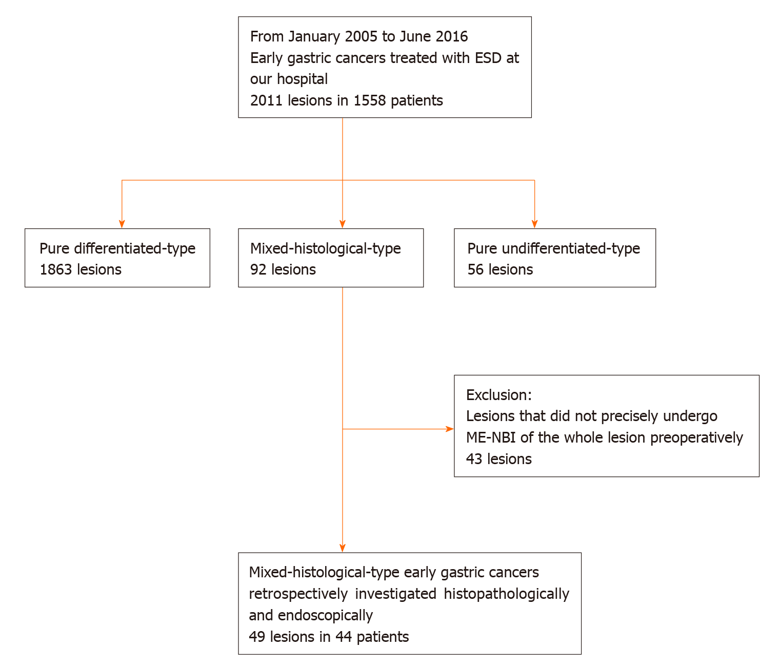
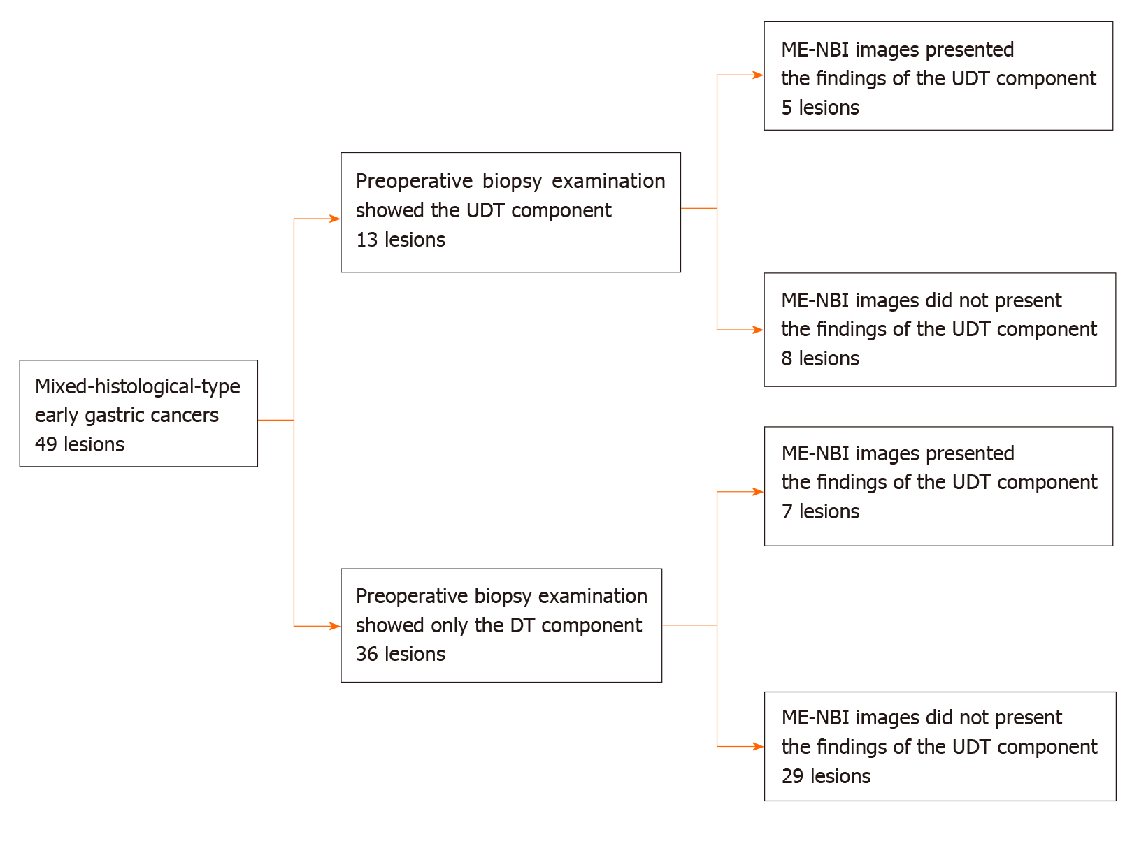
Figure 1 Patient recruitment.
ESD: Endoscopic submucosal dissection; ME-NBI: Magnifying endoscopy with narrow-band imaging.
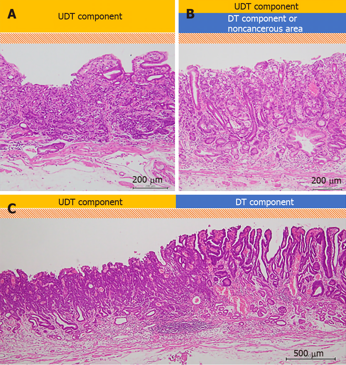
Figure 2 Evaluation of histopathological examination (hematoxylin-eosin staining).
A schema of the three histopathological findings investigated in our study. The scale bars show 200 μm in (A) and (B), and 500 μm in (C). A: The whole mucosal layer occupation of undifferentiated-type (UDT) component (100 ×); B: Exposure of UDT component to the mucosal surface (100 ×); C: Clear border between differentiated-type and UDT components (40 ×). DT: Differentiated-type; UDT: Undifferentiated-type.
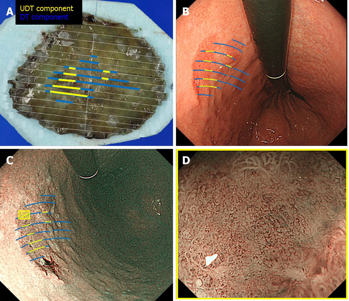
Figure 3 Correlation between pathology and endoscopic findings.
Preoperative endoscopic images were reviewed to determine whether the endoscopic finding of undifferentiated-type (UDT) component was recognizable within the area where UDT component was present in the histopathological examination. A: Endoscopically resected specimen cut into 2-mm-thick slices with mapping according to the histological types. Blue lines indicate differentiated-type component, while yellow lines indicate UDT component; B: Endoscopic images upon white light imaging onto which the histological type is mapped; C: Endoscopic images upon narrow-band imaging onto which the histological type is mapped; D: Magnifying endoscopy with narrow-band imaging finding of the designated UDT area with the yellow square in Figure 3C. ME-NBI: Magnifying endoscopy with narrow-band imaging; DT: Differentiated-type; UDT: Undifferentiated-type.
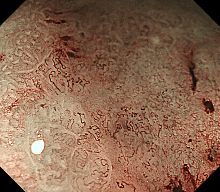
Figure 4 A representative magnifying endoscopy with narrow-band imaging image of the undifferentiated-type component.
Magnifying endoscopy with narrow-band imaging finding of undifferentiated-type. Component was defined as satisfying both a microvascular pattern, which has an isolated and disordered quality and a microstructure pattern, which lacks visibility or even disappears. It complied with the finding described as a corkscrew pattern.
Figure 5 Diagnostic accuracy of pretreatment biopsy examination for undifferentiated-type-component and possible additional effect of magnifying endoscopy with narrow-band imaging.
DT: Differentiated-type; UDT: Undifferentiated-type; ME-NBI: Magnifying endoscopy with narrow-band imaging.
- Citation: Ozeki Y, Hirasawa K, Kobayashi R, Sato C, Tateishi Y, Sawada A, Ikeda R, Nishio M, Fukuchi T, Makazu M, Taguri M, Maeda S. Histopathological validation of magnifying endoscopy for diagnosis of mixed-histological-type early gastric cancer. World J Gastroenterol 2020; 26(36): 5450-5462
- URL: https://www.wjgnet.com/1007-9327/full/v26/i36/5450.htm
- DOI: https://dx.doi.org/10.3748/wjg.v26.i36.5450


