Copyright
©The Author(s) 2020.
World J Gastroenterol. Sep 7, 2020; 26(33): 4945-4959
Published online Sep 7, 2020. doi: 10.3748/wjg.v26.i33.4945
Published online Sep 7, 2020. doi: 10.3748/wjg.v26.i33.4945
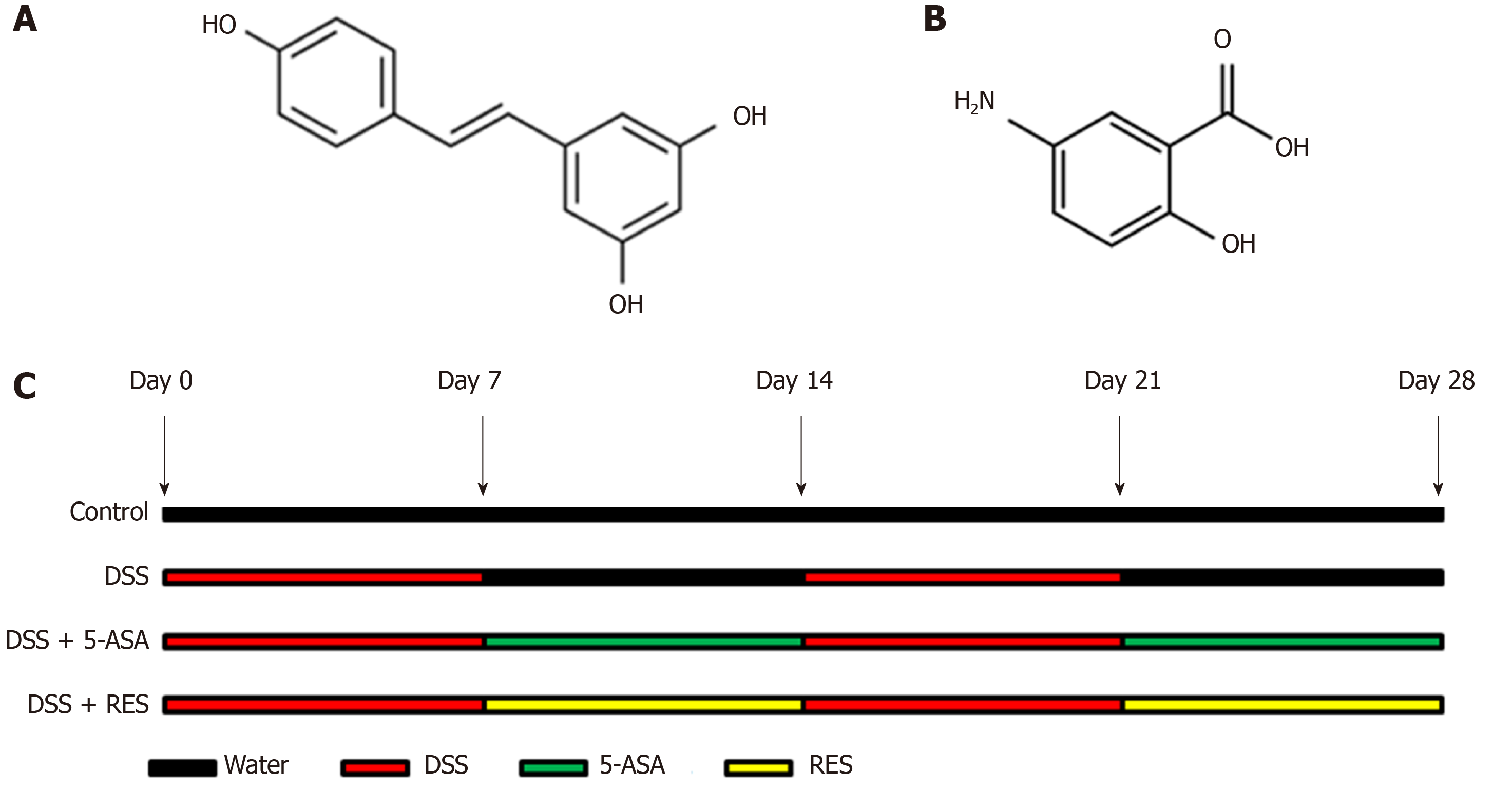
Figure 1 Chemical structures of resveratrol and 5-aminosalicylic acid, and timeline of in vivo experiment.
A: The chemical structures of resveratrol; B: The chemical structures of 5-aminosalicylic acid; and C: Timeline of in vivo experiment. Dextran sulfate sodium (DSS) group was induced by two cycles of intake of drinking water containing 3% DSS for 7 d and normal drinking water for 7 d. Other groups were also treated by two cycles, first received DSS by drinking water for 7 d, followed by treatment of 5-aminosalicylic acid or resveratrol by gavage for 7 d. DSS: Dextran sulfate sodium; 5-ASA: 5-aminosalicylic acid; RES: Resveratrol.
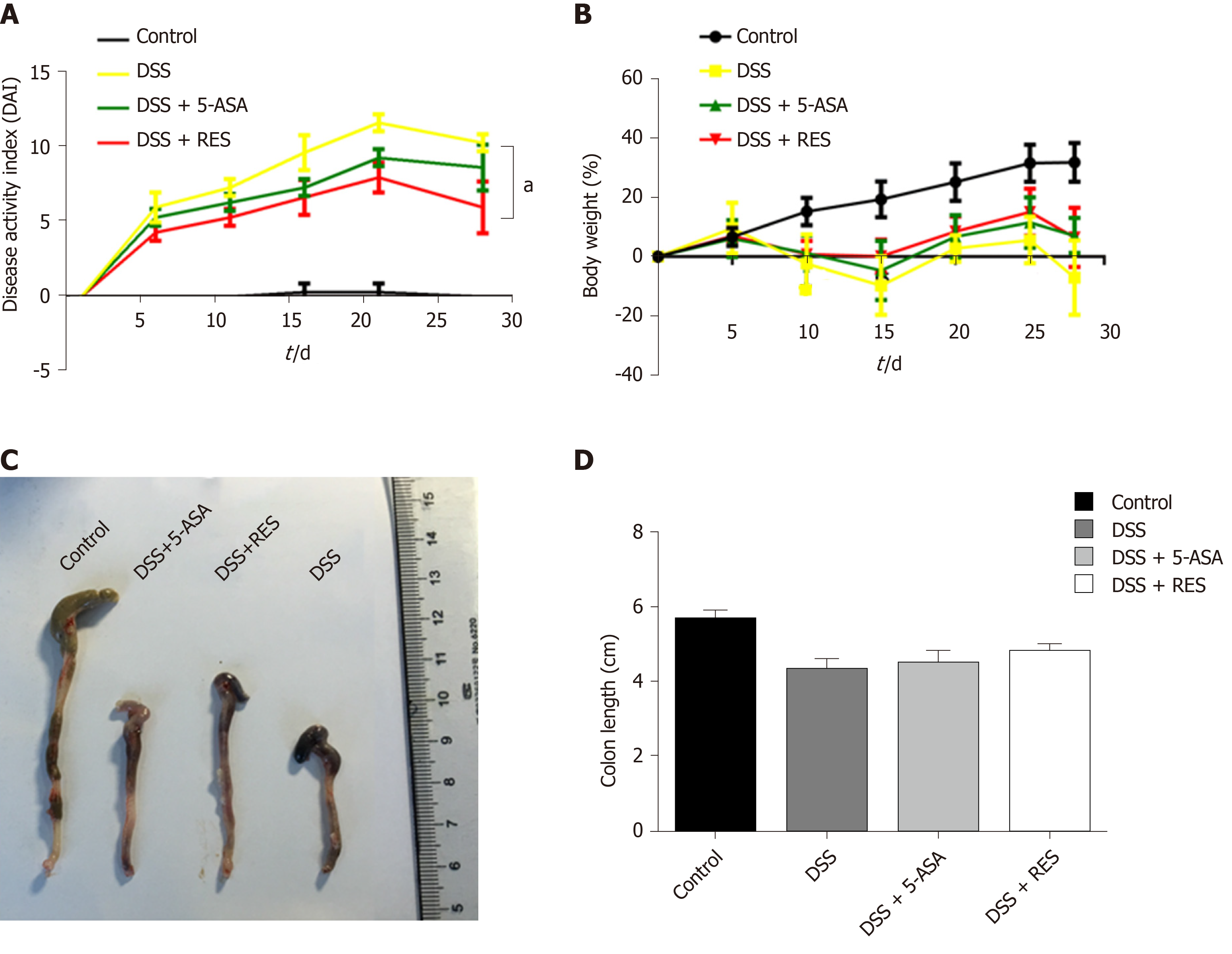
Figure 2 Clinical evaluation of dextran sulfate sodium-induced chronic colitis by disease activity index, body mass loss and colon length (n = 12 for the control group, n = 9 for disease activity index group, n = 11 for dextran sulfate sodium + 5-aminosalicylic acid group, n = 12 for dextran sulfate sodium + resveratrol treatment group).
A: The disease activity index score was reduced in the resveratrol treated group compared with the dextran sulfate sodium group (aP < 0.05); B and C: Resveratrol treatment increased the body mass and colon length of dextran sulfate sodium-induced colitis mice. Body mass loss was calculated as (detected body mass - initial mass)/initial mass. DSS: Dextran sulfate sodium; 5-ASA: 5-aminosalicylic acid; RES: Resveratrol.
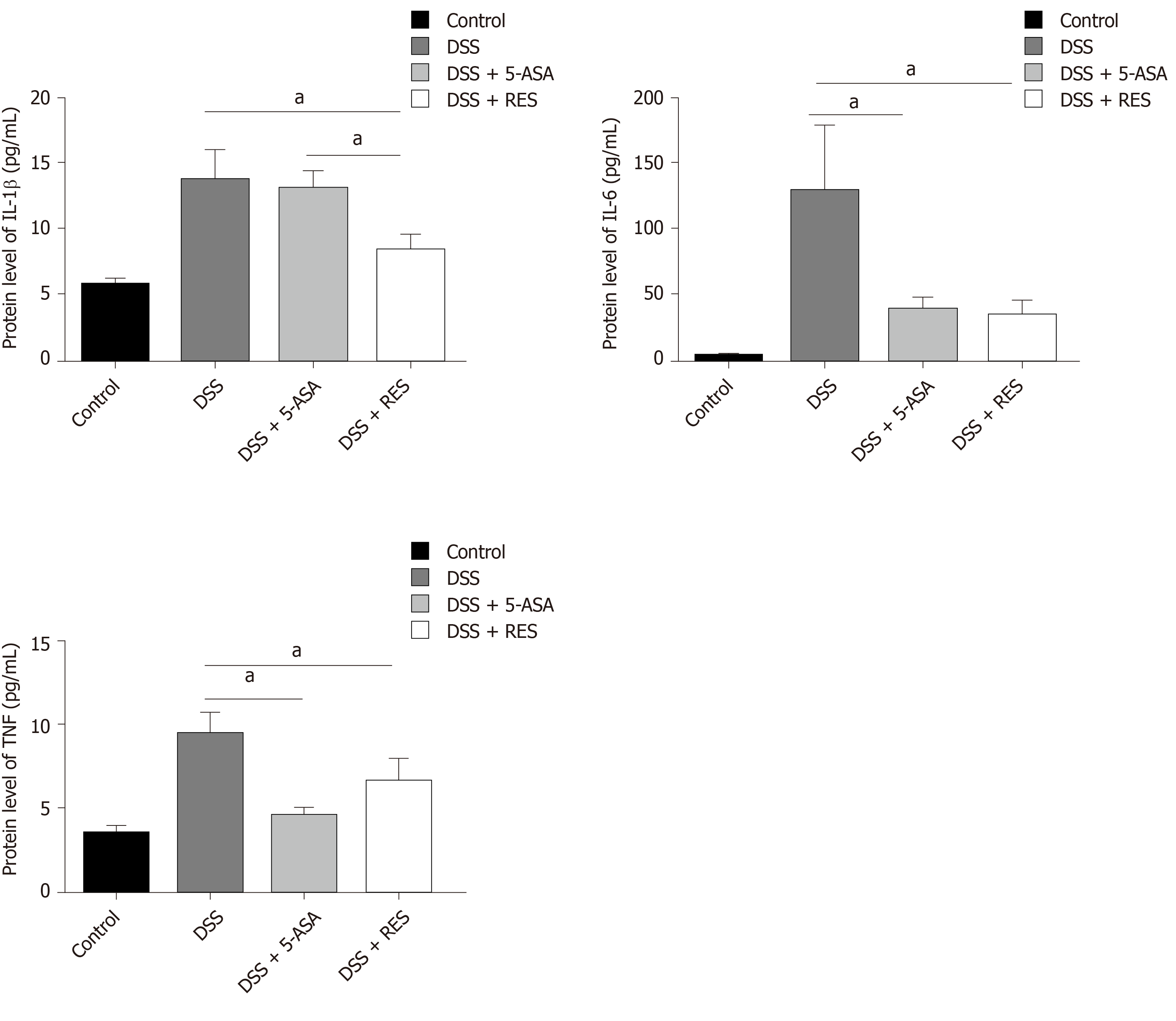
Figure 3 Inflammatory cytokine expression in the dextran sulfate sodium-induced chronic colitis determined by enzyme-linked immunosorbent assay.
The levels of interleukin-1β, interleukin-6 and tumor necrosis factor-α were decreased in the resveratrol treatment group compared with the dextran sulfate sodium group. aP < 0.05. TNF: Tumor necrosis factor; DSS: Dextran sulfate sodium; 5-ASA: 5-aminosalicylic acid; RES: Resveratrol.
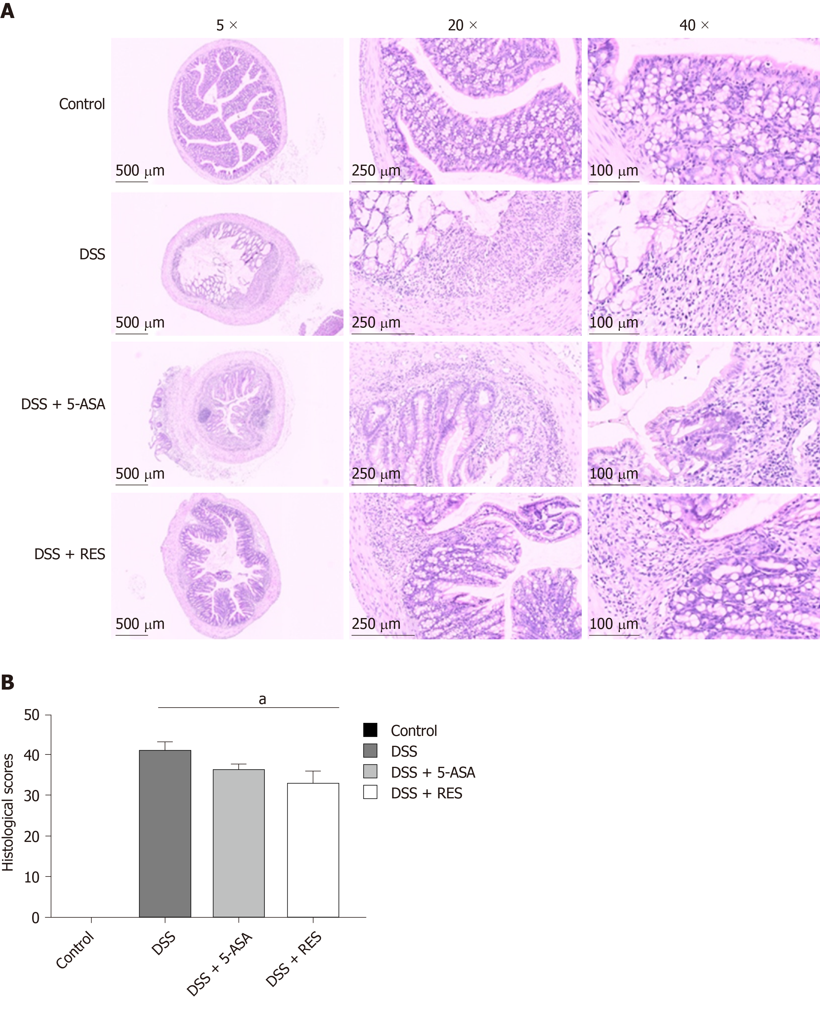
Figure 4 Histological staining showed colitis induced dysfunction.
A: Dextran sulfate sodium group showed significant colonic mucosal damages, crypt depletion, infiltration of inflammatory cells into the mucosa and submucosa, loss of epithelial barrier. Resveratrol treatment group and 5-aminosalicylic acid could alleviate colitis-induced intestinal mucosal barrier dysfunction; and B: The histological score of resveratrol treatment group was lower than the dextran sulfate sodium group. aP < 0.05. DSS: Dextran sulfate sodium; 5-ASA: 5-aminosalicylic acid; RES: Resveratrol.
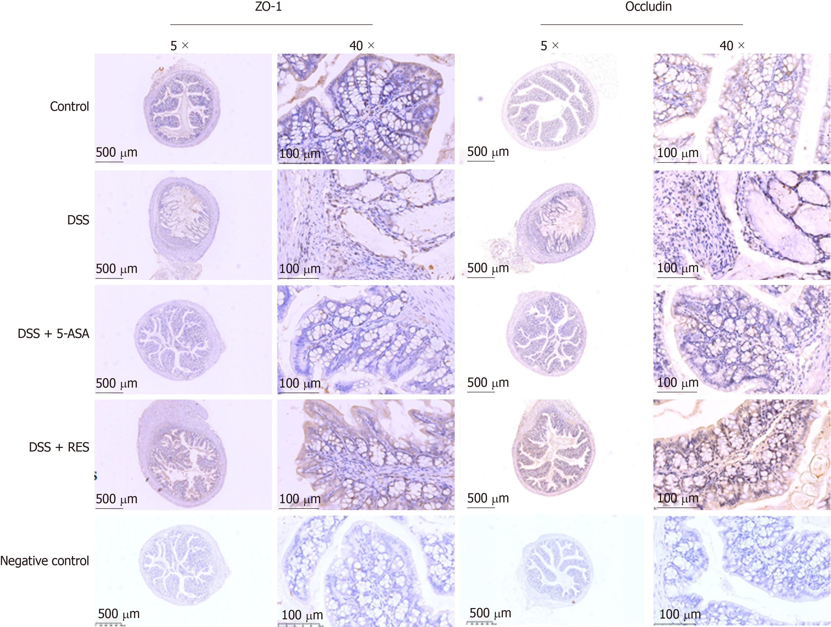
Figure 5 Immunohistochemical staining of ZO-1 and occludin.
Expressions of ZO-1 and occludin were higher in resveratrol treatment than in the dextran sulfate sodium group and 5-aminosalicylic acid treated group. Negative control: antibody was replaced by phosphate buffer saline. DSS: Dextran sulfate sodium; 5-ASA: 5-aminosalicylic acid; RES: Resveratrol.
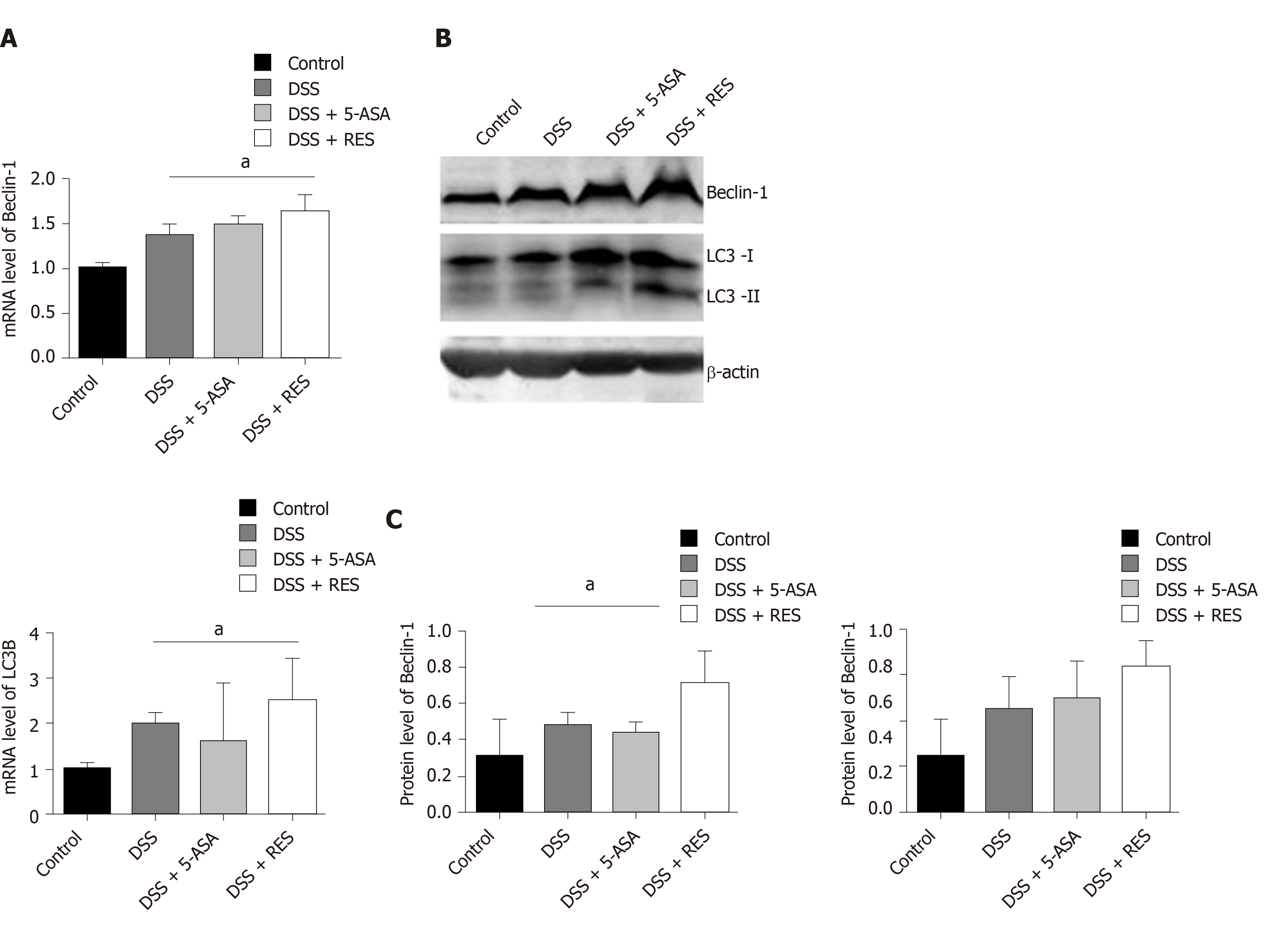
Figure 6 Expression levels of Beclin-1 and LC3B in dextran sulfate sodium induced chronic colitis.
A: A substantial increase in the messenger ribonucleic acid expression level of LC3B and Beclin-1 was observed in the dextran sulfate sodium (DSS) + resveratrol treatment group compared with the DSS group (P < 0.05); B and C: The Western blotting showed that resveratrol treatment induced significant increases in the LC3-II/I ratio and Beclin-1 level in DSS-induced colitis mice; Mean grey level of Beclin-1 and LC3B protein level was increased in DSS + resveratrol treatment group. aP < 0.05. DSS: Dextran sulfate sodium; 5-ASA: 5-aminosalicylic acid; mRNA: Messenger ribonucleic acid; RES: Resveratrol.
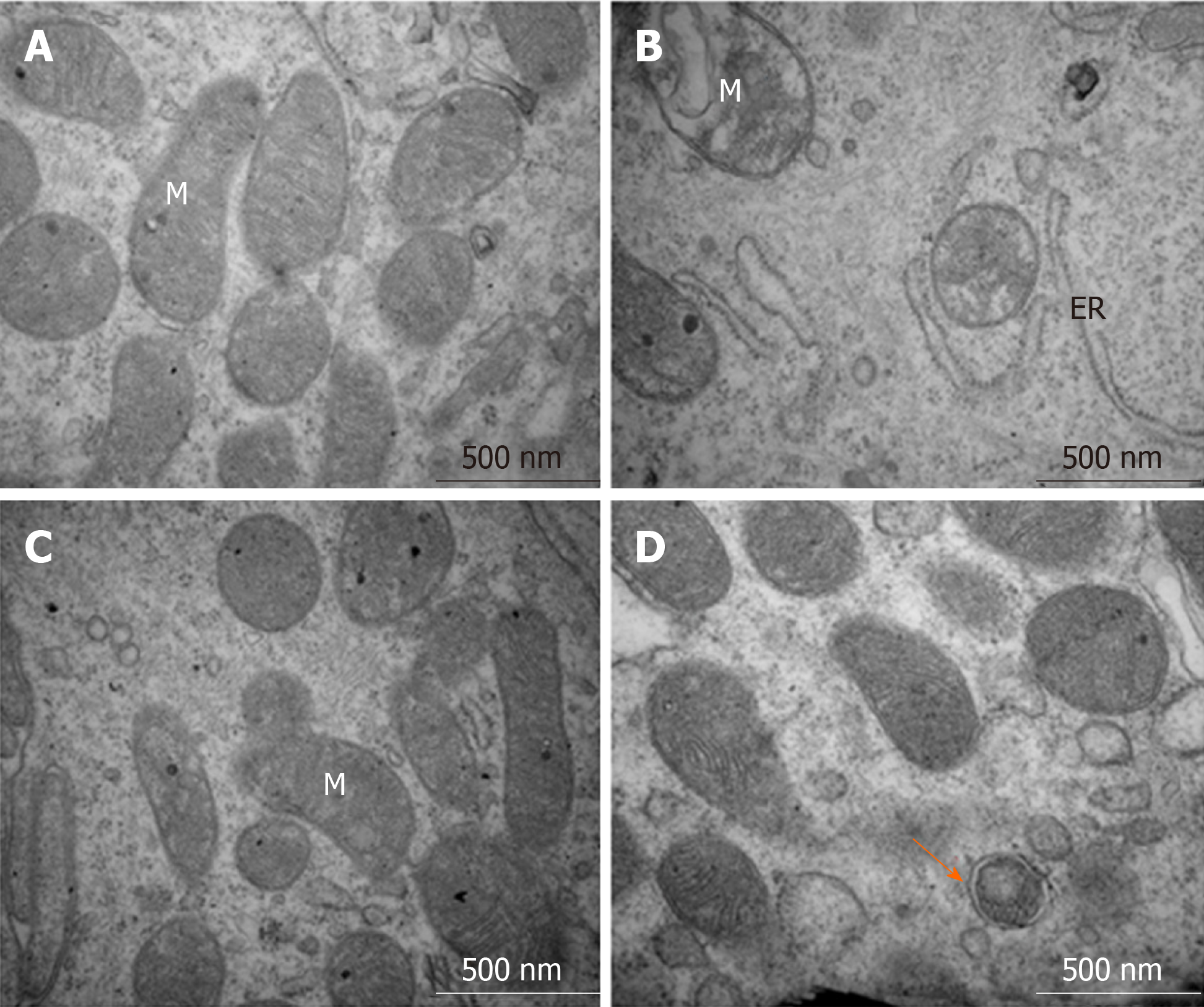
Figure 7 Structures of the intestinal epithelial cells and autolysosomes observed by transmission electron microscopy.
A: Control group showed normal organelle structure; B: Dextran sulfate sodium induced colitis group showed mitochondrial swelling and ruptures of internal cristae, along with distention of the rough endoplasmic reticulum; C: 5-aminosalicylic acid group revealed that mitochondrial swelling was not obvious, and mitochondrial cristae showed blurred appearance; and D: Resveratrol treatment group showed that organelle structure was basically normal, and autophagosome could be observed. ER: Endoplasmic reticulum, M: Mitochondria.
- Citation: Pan HH, Zhou XX, Ma YY, Pan WS, Zhao F, Yu MS, Liu JQ. Resveratrol alleviates intestinal mucosal barrier dysfunction in dextran sulfate sodium-induced colitis mice by enhancing autophagy. World J Gastroenterol 2020; 26(33): 4945-4959
- URL: https://www.wjgnet.com/1007-9327/full/v26/i33/4945.htm
- DOI: https://dx.doi.org/10.3748/wjg.v26.i33.4945


