Copyright
©The Author(s) 2020.
World J Gastroenterol. Jul 14, 2020; 26(26): 3834-3850
Published online Jul 14, 2020. doi: 10.3748/wjg.v26.i26.3834
Published online Jul 14, 2020. doi: 10.3748/wjg.v26.i26.3834
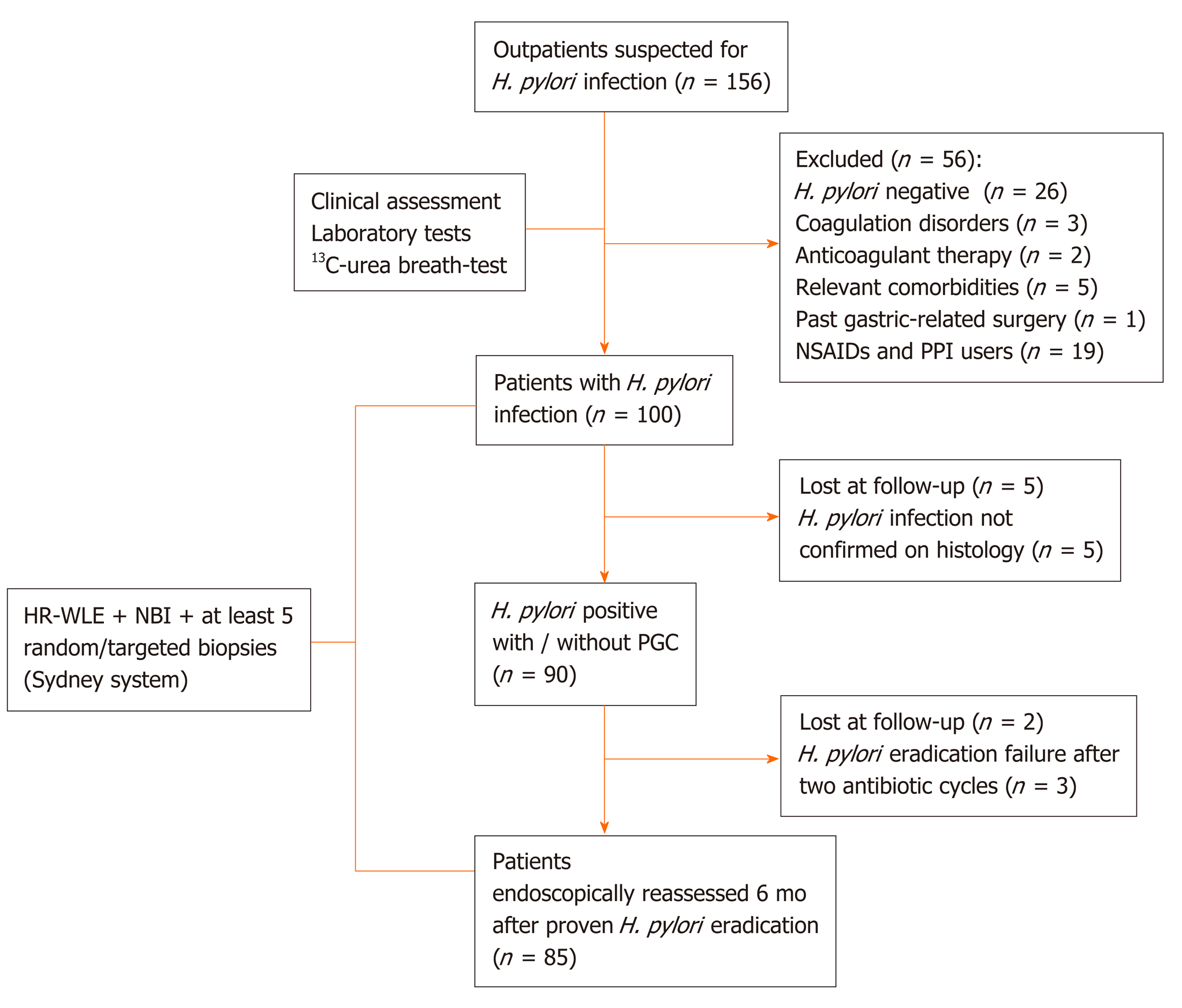
Figure 1 Flow diagram of patient’s selection.
H. pylori: Helicobacter pylori; NSAIDs: Non-steroidal anti-inflammatory drugs; PPI: Proton pump inhibitor; HR-WLE: High resolution-white light endoscopy; NBI: Narrow band imaging; PGC: Precancerous gastric conditions.
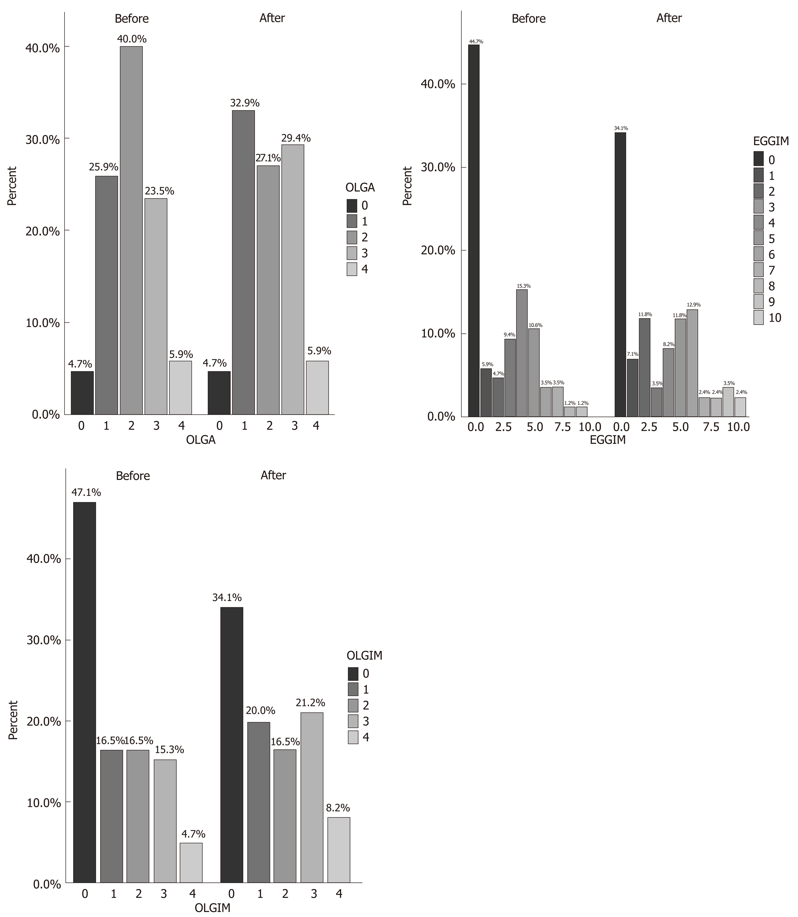
Figure 2 Prevalence of Operative Link on Gastritis Assessment, endoscopic grading of gastric intestinal metaplasia, and Operative Link on Gastric Intestinal Metaplasia scores, respectively in the 85 patients before and after Helicobacter pylori eradication.
OLGA: Operative Link on Gastritis Assessment; EGGIM: Endoscopic grading of gastric intestinal metaplasia; OLGIM: Operative Link on Gastric Intestinal Metaplasia.
Figure 3 Gastric white-light endoscopy, narrow band imaging and histological evaluation before Helicobacter pylori eradication.
A, B: Gastritis during white-light endoscopy and narrow band imaging assessment of corpus before Helicobacter pylori eradication; and C: Histological evaluation: moderately atrophic chronically active gastritis with lymphoplasmacellular infiltration of the lamina propria and foveolar epithelium hyperplasia (fundus before Helicobacter pylori eradication. Sections of 3 microns colored with Hematoxylin Eosin and Giemsa respectively. Magnifications: × 10).
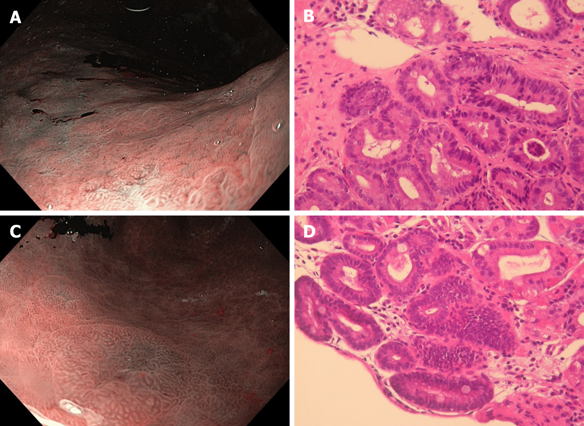
Figure 4 Gastric narrow band imaging and histological evaluation after Helicobacter pylori eradication.
A: Low-grade dysplasia on flat lesion during narrow band imaging assessment of corpus after Helicobacter pylori eradication; B: Histological appearance: inactive chronically mild atrophic gastritis with intestinal metaplasia and low-grade dysplasia on intestinal metaplastic epithelium (fundus after Helicobacter pylori eradication); C: Low-grade dysplasia aspect of antrum on visible lesion during narrow band imaging assessment after Helicobacter pylori eradication; and D: Histological appearance: inactive chronically moderate atrophic gastritis with intestinal metaplasia and low-grade dysplasia on intestinal metaplastic epithelium (antrum after Helicobacter pylori eradication). Hematoxylin Eosin. Sections of 3 microns. Magnification: × 10.
- Citation: Panarese A, Galatola G, Armentano R, Pimentel-Nunes P, Ierardi E, Caruso ML, Pesce F, Lenti MV, Palmitessa V, Coletta S, Shahini E. Helicobacter pylori-induced inflammation masks the underlying presence of low-grade dysplasia on gastric lesions. World J Gastroenterol 2020; 26(26): 3834-3850
- URL: https://www.wjgnet.com/1007-9327/full/v26/i26/3834.htm
- DOI: https://dx.doi.org/10.3748/wjg.v26.i26.3834


