Copyright
©The Author(s) 2020.
World J Gastroenterol. Jul 14, 2020; 26(26): 3767-3779
Published online Jul 14, 2020. doi: 10.3748/wjg.v26.i26.3767
Published online Jul 14, 2020. doi: 10.3748/wjg.v26.i26.3767
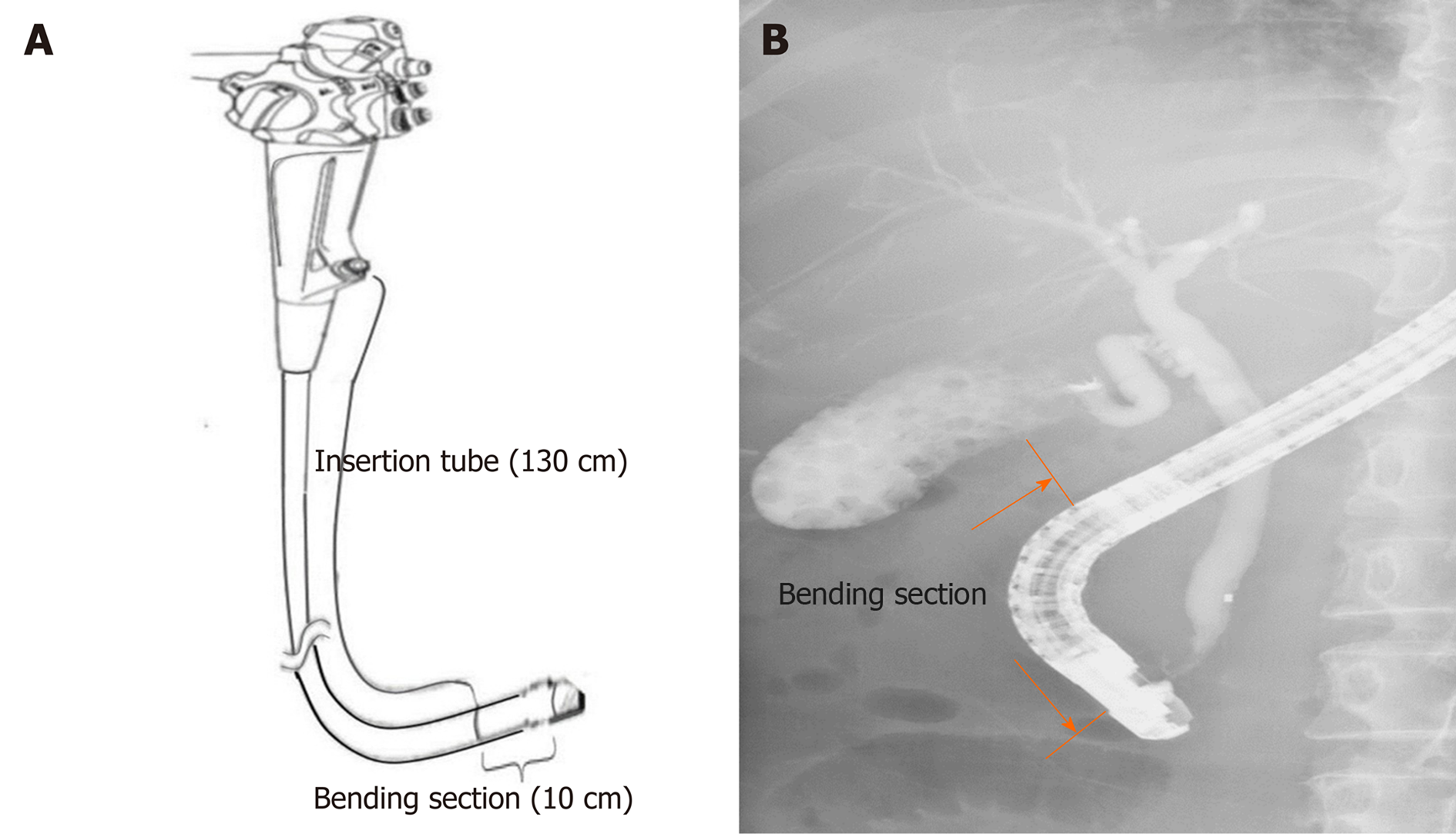
Figure 1 A scheme depicting the working channel of a duodenoscope.
Insertion tube (130 cm) starting from the biopsy channel opening at the proximal part (A) and bending section (10 cm) proximal to the elevator mechanism (B).
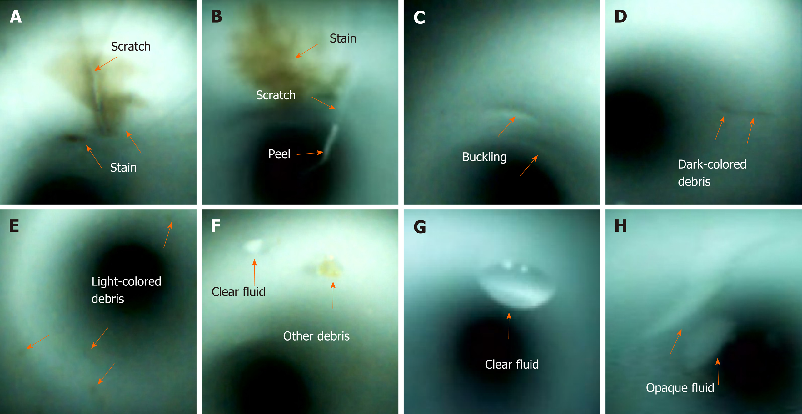
Figure 2 Abnormal visual inspection findings (orange arrow) inside the working channels, including scratches (A and B), scratch with an adherent peel (B), buckling (C), stains (A and B), dark-colored debris (D), light-colored debris (E), other debris (F), clear fluid (F and G), and opaque fluid (H).
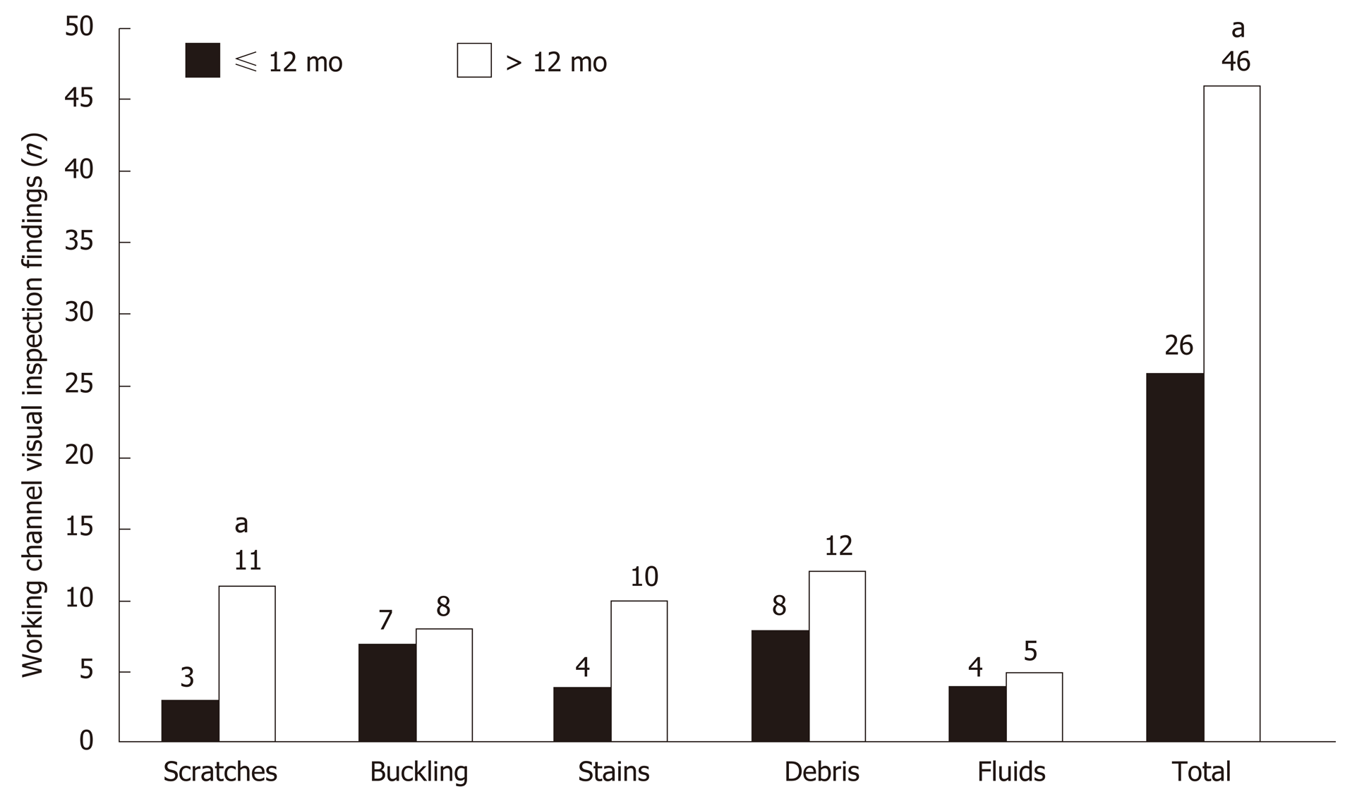
Figure 3 Comparison of visual inspection findings between > 12-mo-old and ≤ 12mo-old duodenoscopes.
aIndicates statistically significant difference.
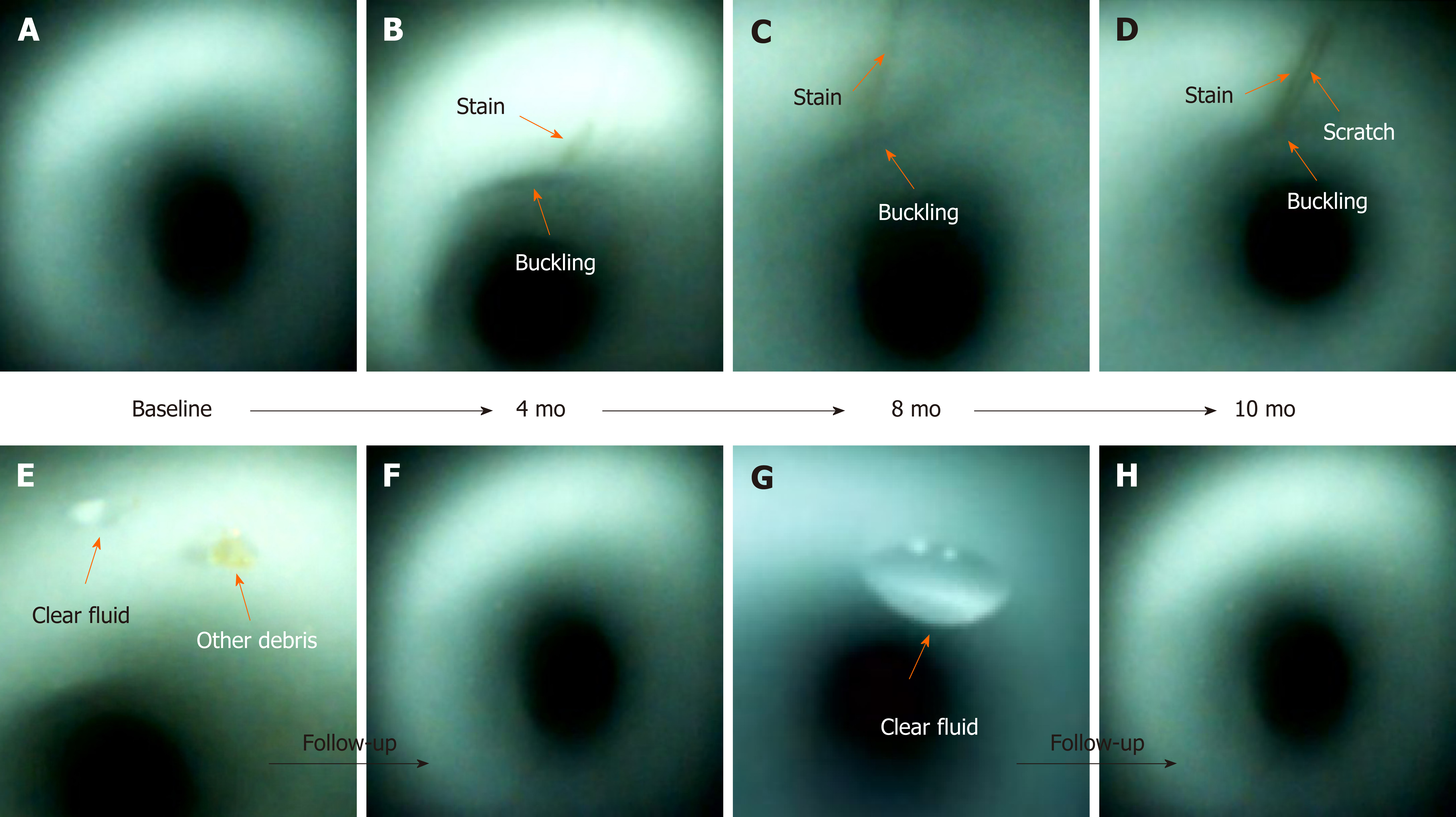
Figure 4 Scratches, buckling, and stains (orange arrow) appeared at the same location inside the working channel during the follow-up inspections (B-D).
Buckling and stains progressively increased in length and width (A-D). A scratch (D) was found. Debris (E and F) and clear fluid (E-H) disappeared after one or more cycles of reprocessing.
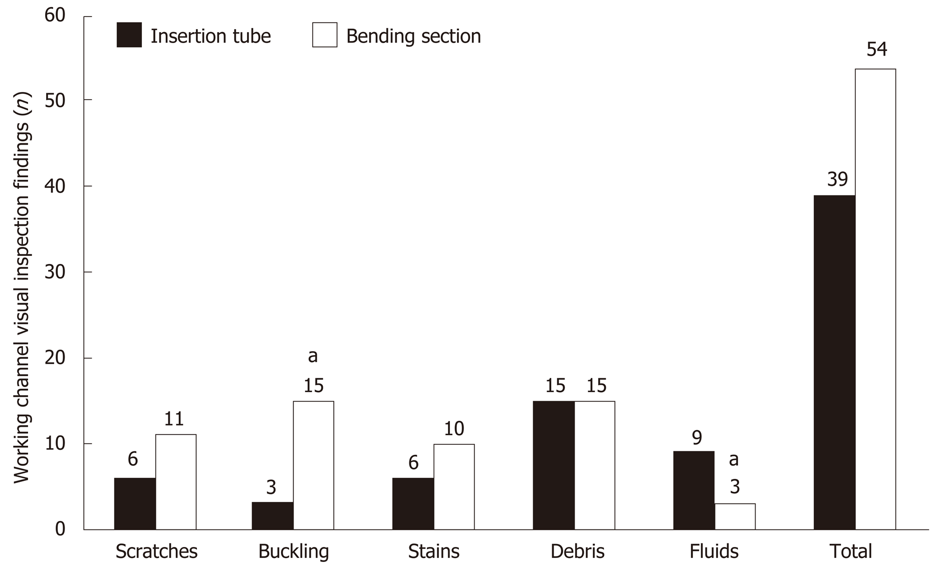
Figure 5 Comparison of visual inspection findings between the bending section and insertion tube.
aIndicates statistically significant difference.
- Citation: Liu TC, Peng CL, Wang HP, Huang HH, Chang WK. SpyGlass application for duodenoscope working channel inspection: Impact on the microbiological surveillance. World J Gastroenterol 2020; 26(26): 3767-3779
- URL: https://www.wjgnet.com/1007-9327/full/v26/i26/3767.htm
- DOI: https://dx.doi.org/10.3748/wjg.v26.i26.3767


