Copyright
©The Author(s) 2020.
World J Gastroenterol. Jun 28, 2020; 26(24): 3432-3446
Published online Jun 28, 2020. doi: 10.3748/wjg.v26.i24.3432
Published online Jun 28, 2020. doi: 10.3748/wjg.v26.i24.3432
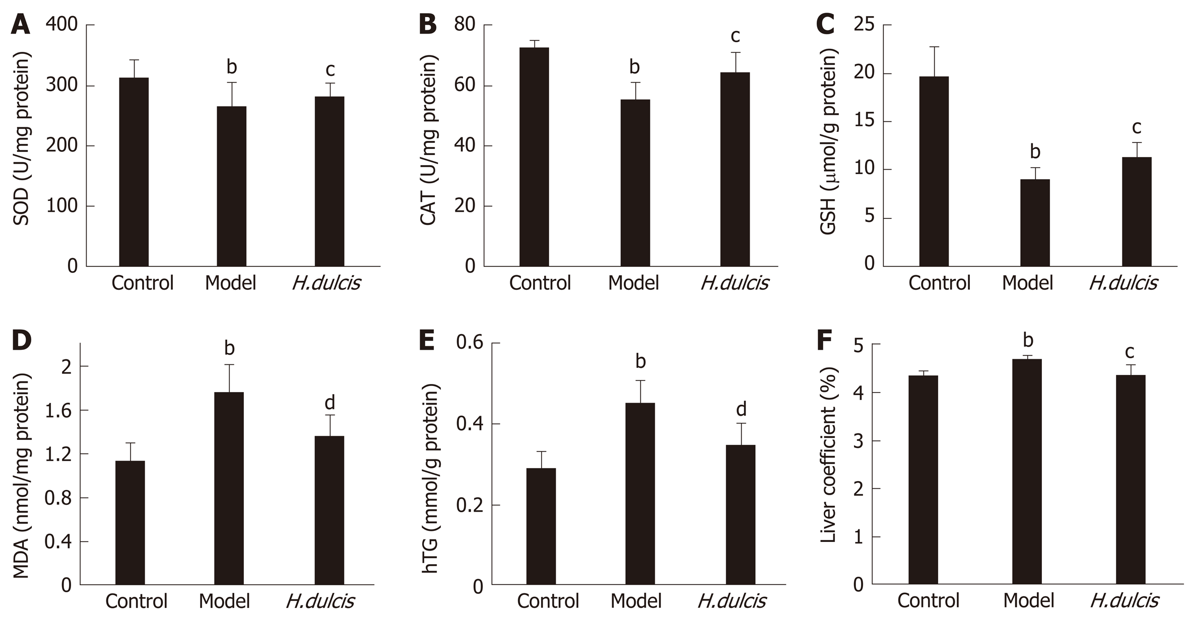
Figure 1 Effects of Hovenia dulcis seeds on hepatic biomarkers in mice with alcoholic liver injury.
A: Superoxide dismutase; B: Catalase; C: Glutathione; D: Malondialdehyde; E: Hepatic triglyceride. aP < 0.05, bP < 0.01 vs control group; cP < 0.05, dP < 0.01 vs model group. Error bars: standard deviations (n = 8). H. dulcis: Hovenia dulcis; SOD: Superoxide dismutase; CAT: Catalase; GSH: Glutathione; MDA: Malondialdehyde; hTG: Hepatic triglyceride.
Figure 2 Histopathological changes (magnified × 400 times, stained by hematoxylin and eosin).
A: Control group; B: Model group; C: Hovenia dulcis seed group. Scale bar: 50 μm; Oval: Area with lipid droplets; Arrow: Inflammatory cells.
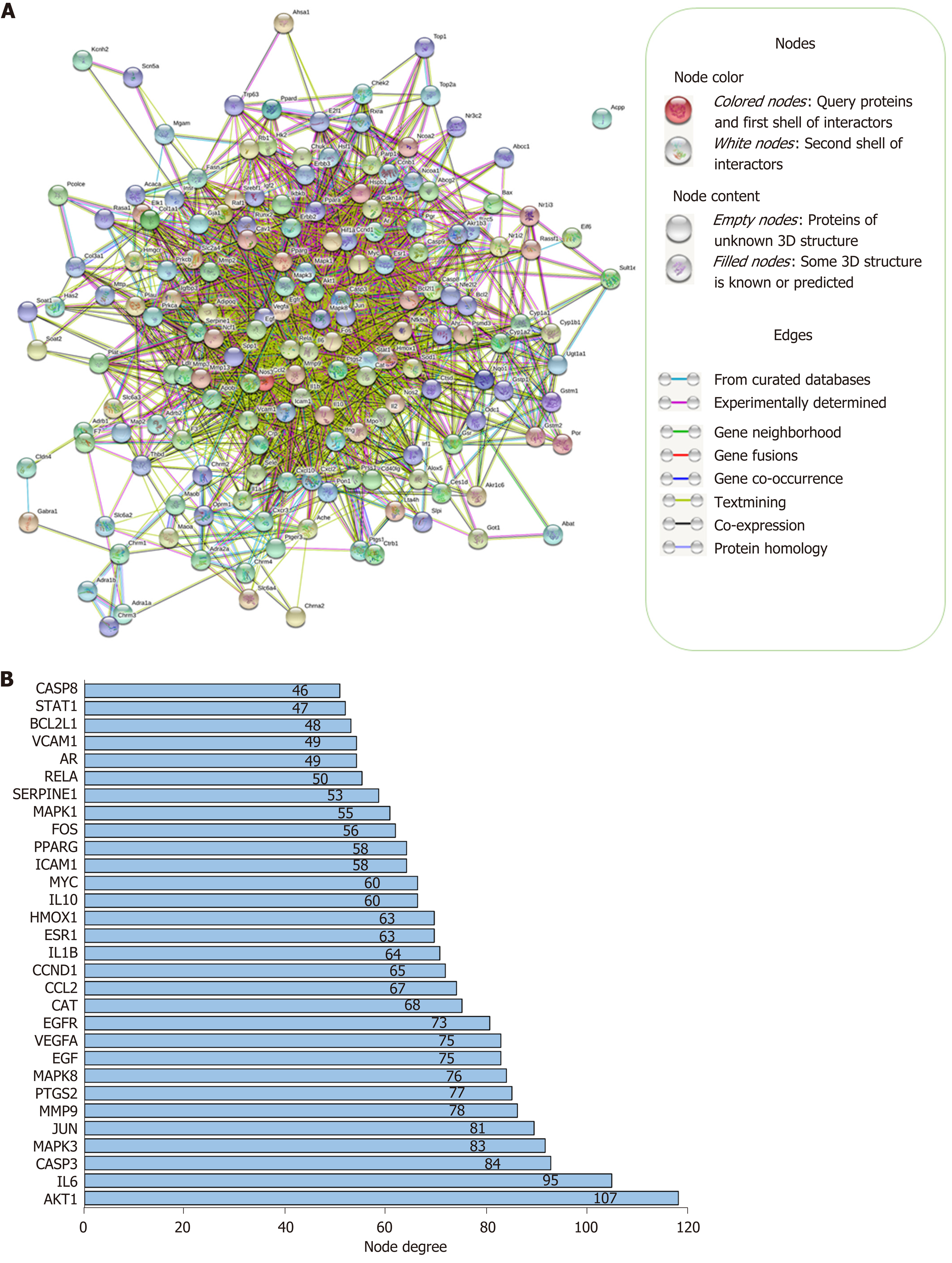
Figure 3 Protein–protein interaction network and node degrees.
A: Interaction of 173 common target proteins; B: Thirty targets with the highest node degrees.
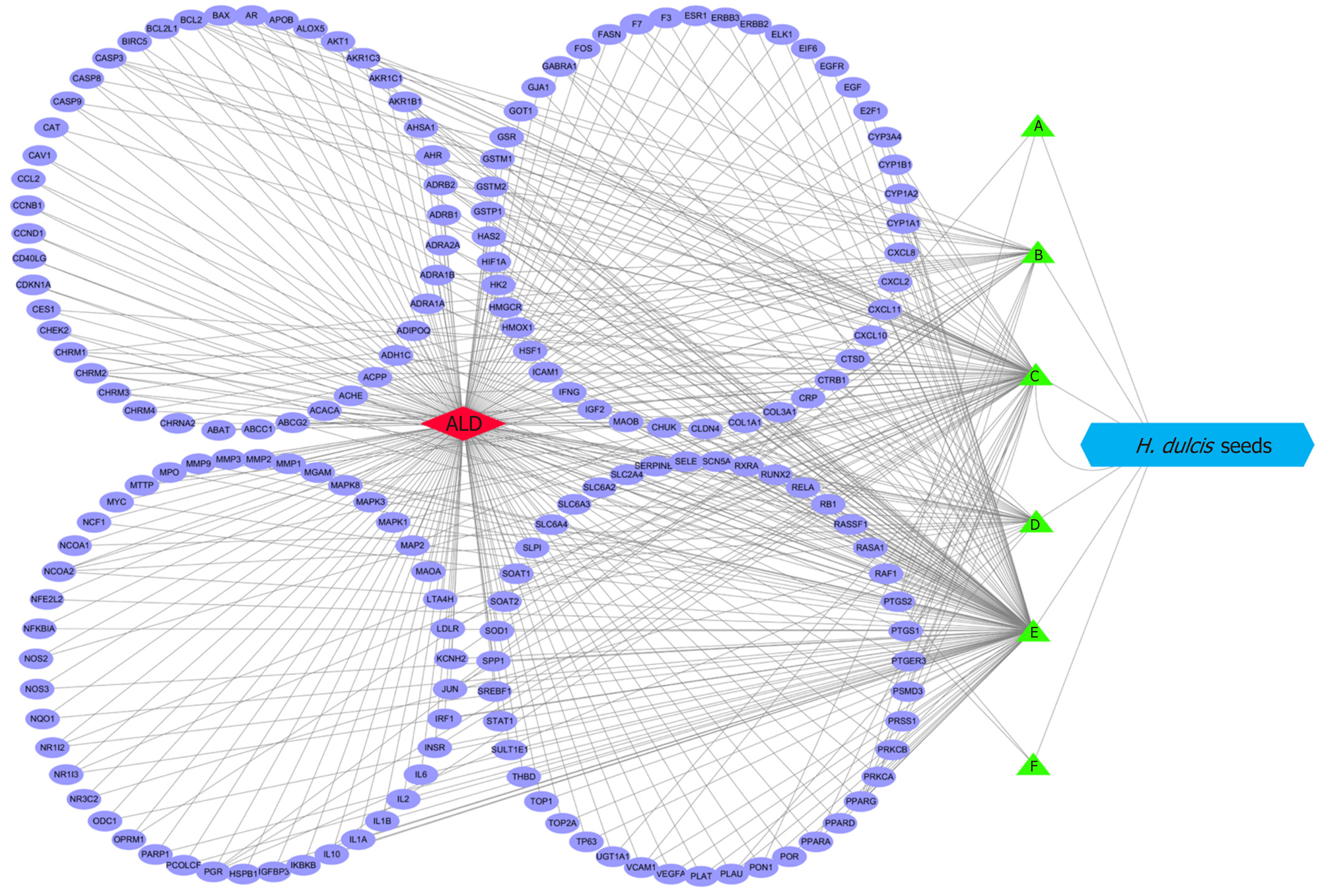
Figure 4 Complex interaction network between targets, the eligible ingredients, and alcoholic liver disease.
A: Perlolyrine; B: β-sitosterol; C: Kaempferol; D: Naringenin; E: Stigmasterol; F: Quercetin. ALD: Alcoholic liver disease; H. dulcis: Hovenia dulcis.
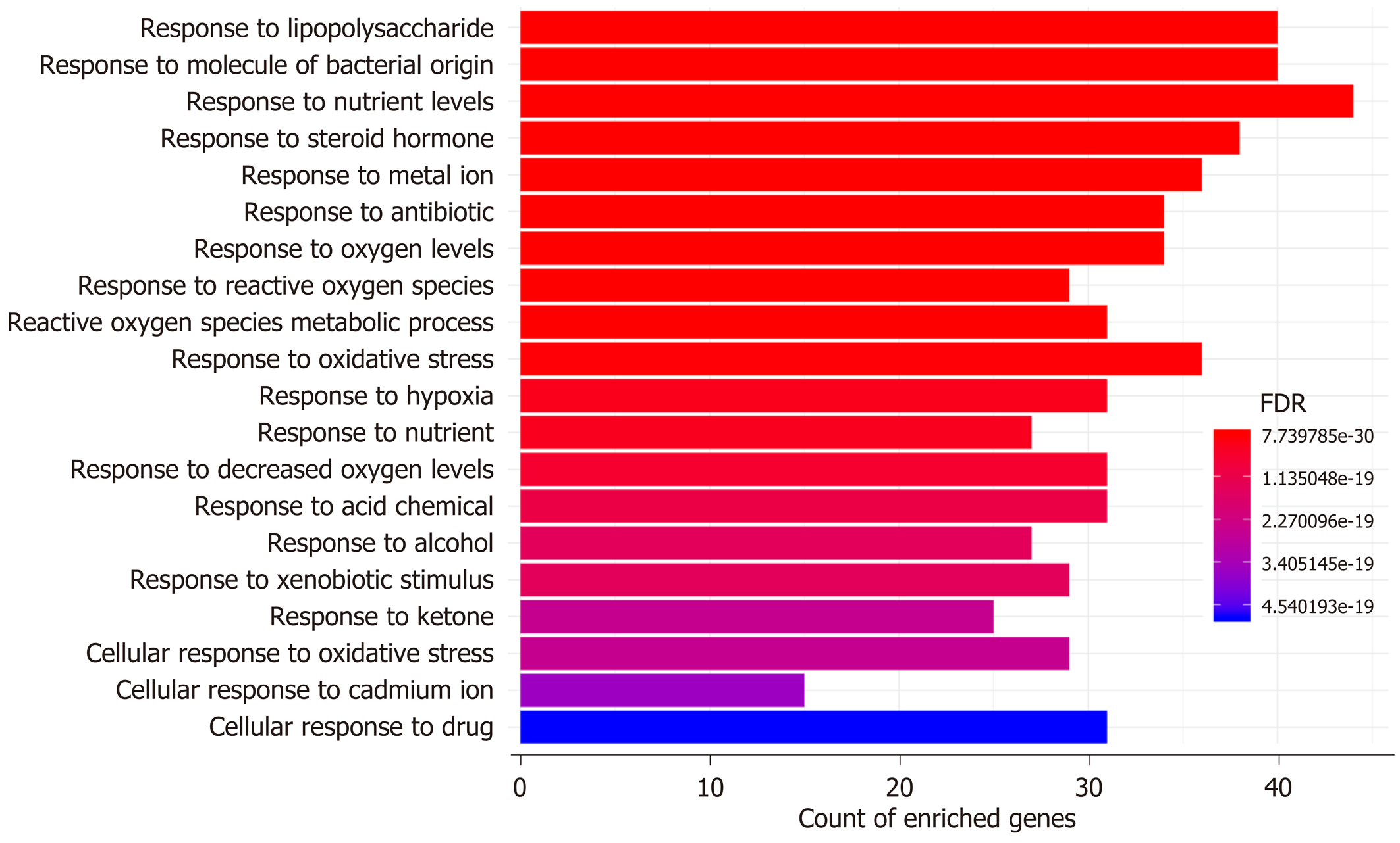
Figure 5 Top 20 enriched biological processes from gene ontology analysis.
FDR: False discovery rate.
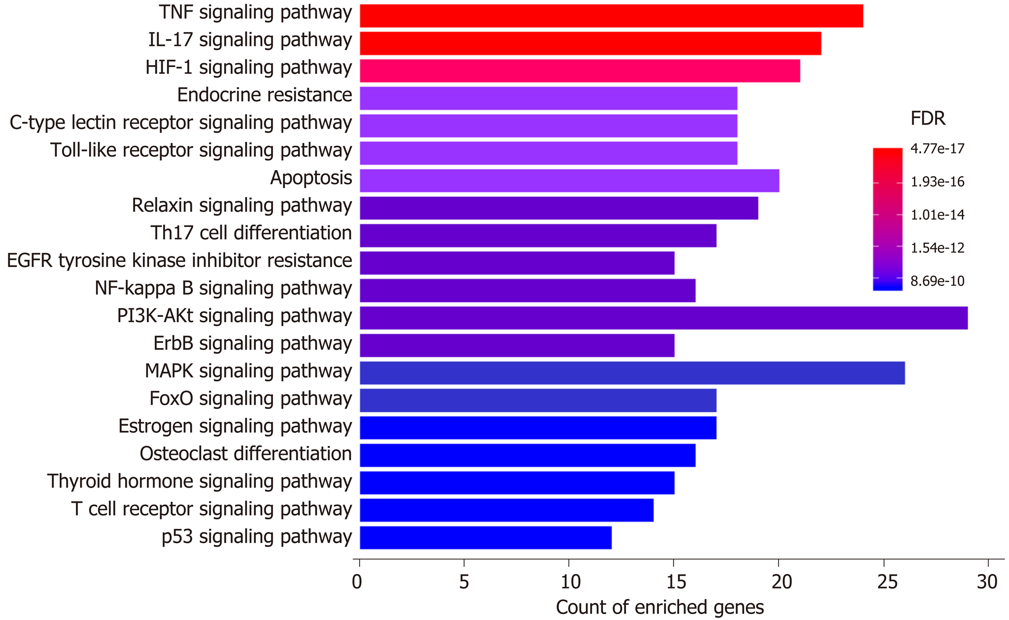
Figure 6 Top 20 enriched pathways from Kyoto Encyclopedia of Genes and Genomes analysis.
FDR: False discovery rate.
- Citation: Meng X, Tang GY, Zhao CN, Liu Q, Xu XY, Cao SY. Hepatoprotective effects of Hovenia dulcis seeds against alcoholic liver injury and related mechanisms investigated via network pharmacology. World J Gastroenterol 2020; 26(24): 3432-3446
- URL: https://www.wjgnet.com/1007-9327/full/v26/i24/3432.htm
- DOI: https://dx.doi.org/10.3748/wjg.v26.i24.3432


