Copyright
©The Author(s) 2020.
World J Gastroenterol. Jun 14, 2020; 26(22): 3012-3023
Published online Jun 14, 2020. doi: 10.3748/wjg.v26.i22.3012
Published online Jun 14, 2020. doi: 10.3748/wjg.v26.i22.3012
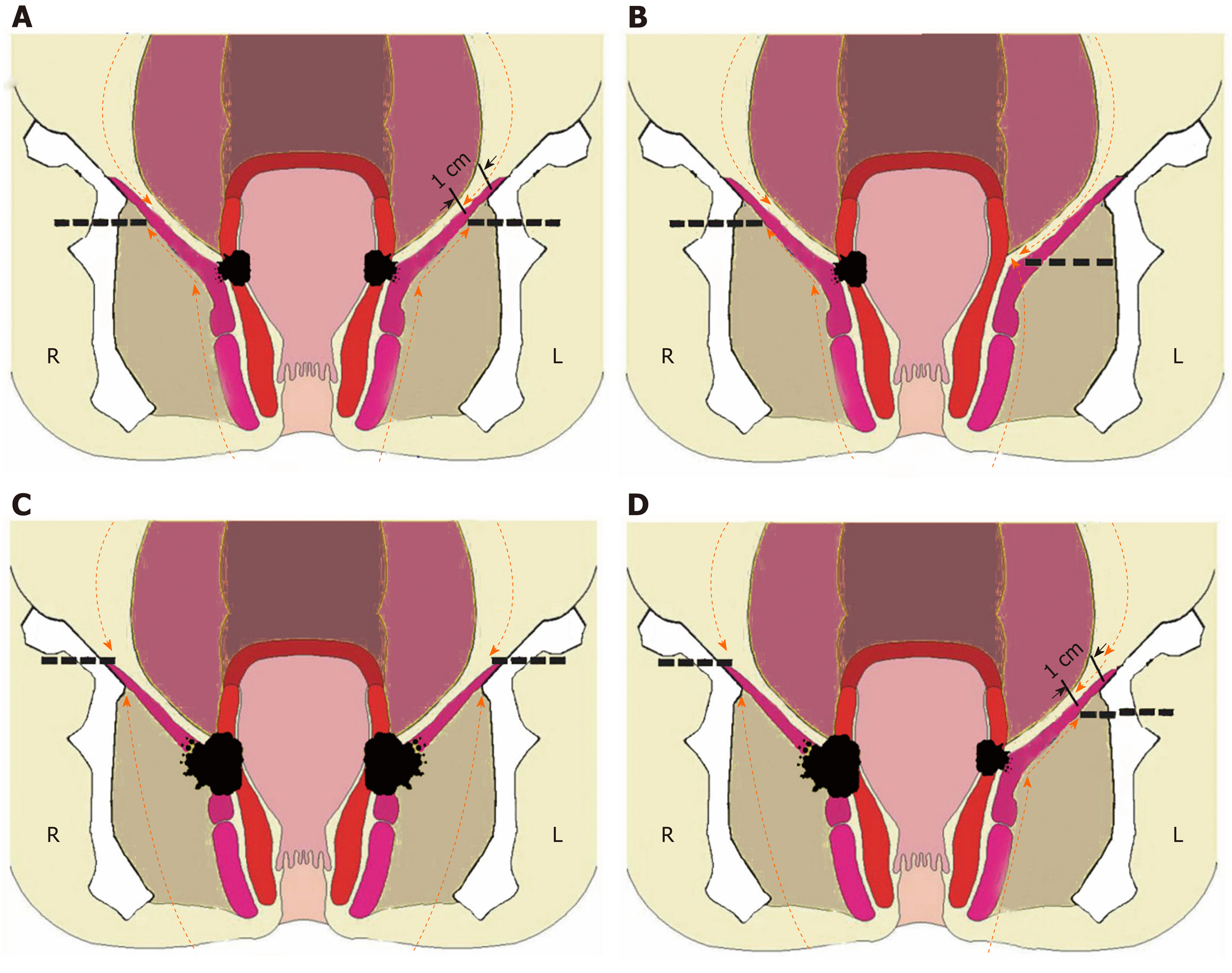
Figure 1 Individualized extralevator abdominoperineal excision procedure.
A: Tumor not involving the ischioanal fat or levator ani muscle (T3), leave 1 cm of the levator ani muscles on the pelvic sidewall; B: Tumor located at one side (T3), levator ani muscle on the other side may be left; C: Tumor penetrating the levator ani muscle (T4) bilaterally, dissection should include the fat of the ischioanal fossa and the intact levator ani muscle bilaterally; D: Tumor penetrating the levator ani muscle (T4) unilaterally, part of the ischioanal fat and intact lavator ani muscle should be dissected unilaterally. This Figure is reprinted with authors’permission[13,60].
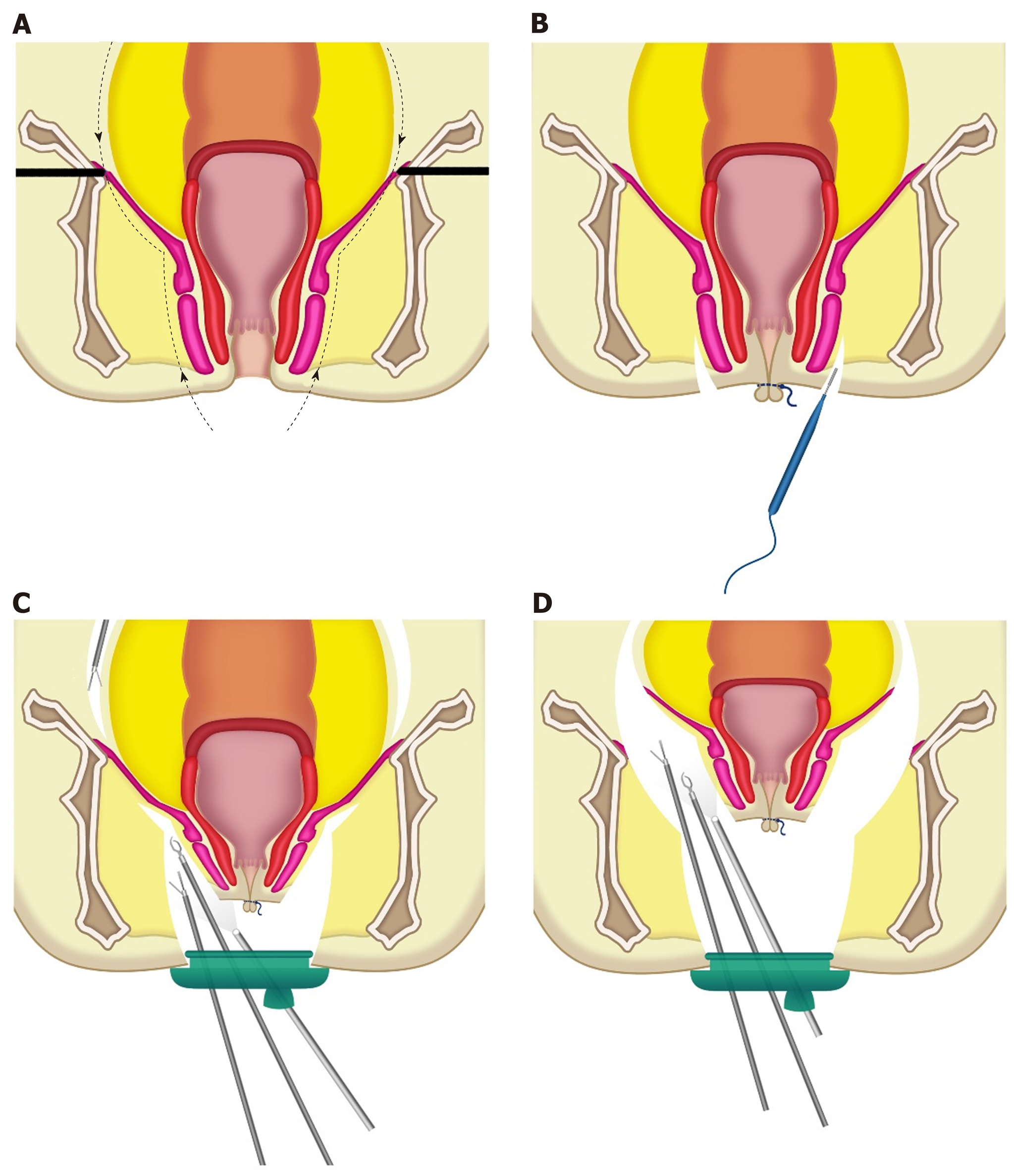
Figure 2 Trans-perineal minimally invasive approach for extralevator abdominoperineal excision procedure.
A: The resection line of transperineal extralevator abdominoperineal excision; B: The anus was closed with a purse-string suture and an incision was made around the anus; C: The dissection was continued outside the external anal sphincter and levator muscle by using the trans-perineal trans-anal minimally invasive surgery (TAMIS) platform. The abdominal procedure was performed at the same time; D: The levator muscles were divided at the lateral most aspect by using the trans-perineal TAMIS platform. Reprinted with permission from the authors[63].
- Citation: Tao Y, Han JG, Wang ZJ. Extralevator abdominoperineal excision for advanced low rectal cancer: Where to go. World J Gastroenterol 2020; 26(22): 3012-3023
- URL: https://www.wjgnet.com/1007-9327/full/v26/i22/3012.htm
- DOI: https://dx.doi.org/10.3748/wjg.v26.i22.3012


