Copyright
©The Author(s) 2020.
World J Gastroenterol. May 7, 2020; 26(17): 2111-2118
Published online May 7, 2020. doi: 10.3748/wjg.v26.i17.2111
Published online May 7, 2020. doi: 10.3748/wjg.v26.i17.2111
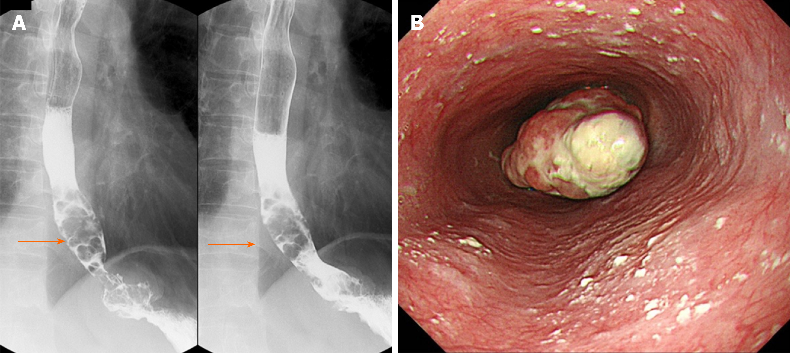
Figure 1 Upper gastrointestinal series and upper gastrointestinal endoscopy.
A: Upper gastrointestinal series reveals stenosis due to the protruding lesion in the lower esophagus (orange arrow); B: Type 1 and 0-IIc lesions at from 35 cm from the incisors toward the esophago-gastric junction.
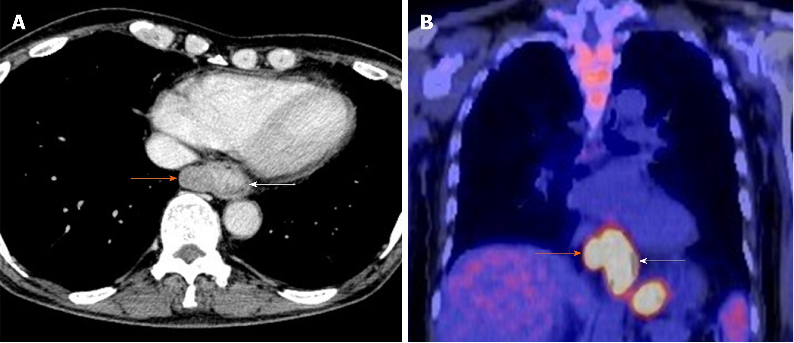
Figure 2 Enhanced computerized tomography and 18F-2-fluoro-2-deoxy-D-glucose positron-emission tomography.
A: Enhanced computerized tomography; B: 18F-2-fluoro-2-deoxy-D-glucose positron-emission tomography. The major axis of the main tumor was 42 mm and maximum standardized uptake value of the lesion was 13.9 at the white arrow. There suspected paraesophageal lymph node metastasis is indicated by the orange arrow.
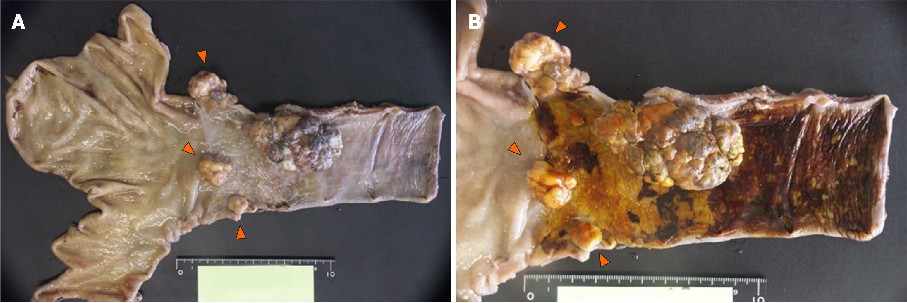
Figure 3 Macroscopic findings.
A: Three polypoid type lesions (orange arrowheads) were identified in the abdominal esophagus in addition to the main 60 mm × 40 mm lesion in the lower esophagus; B: Iodine staining; an unstained zone was detected at the base of the main tumor.
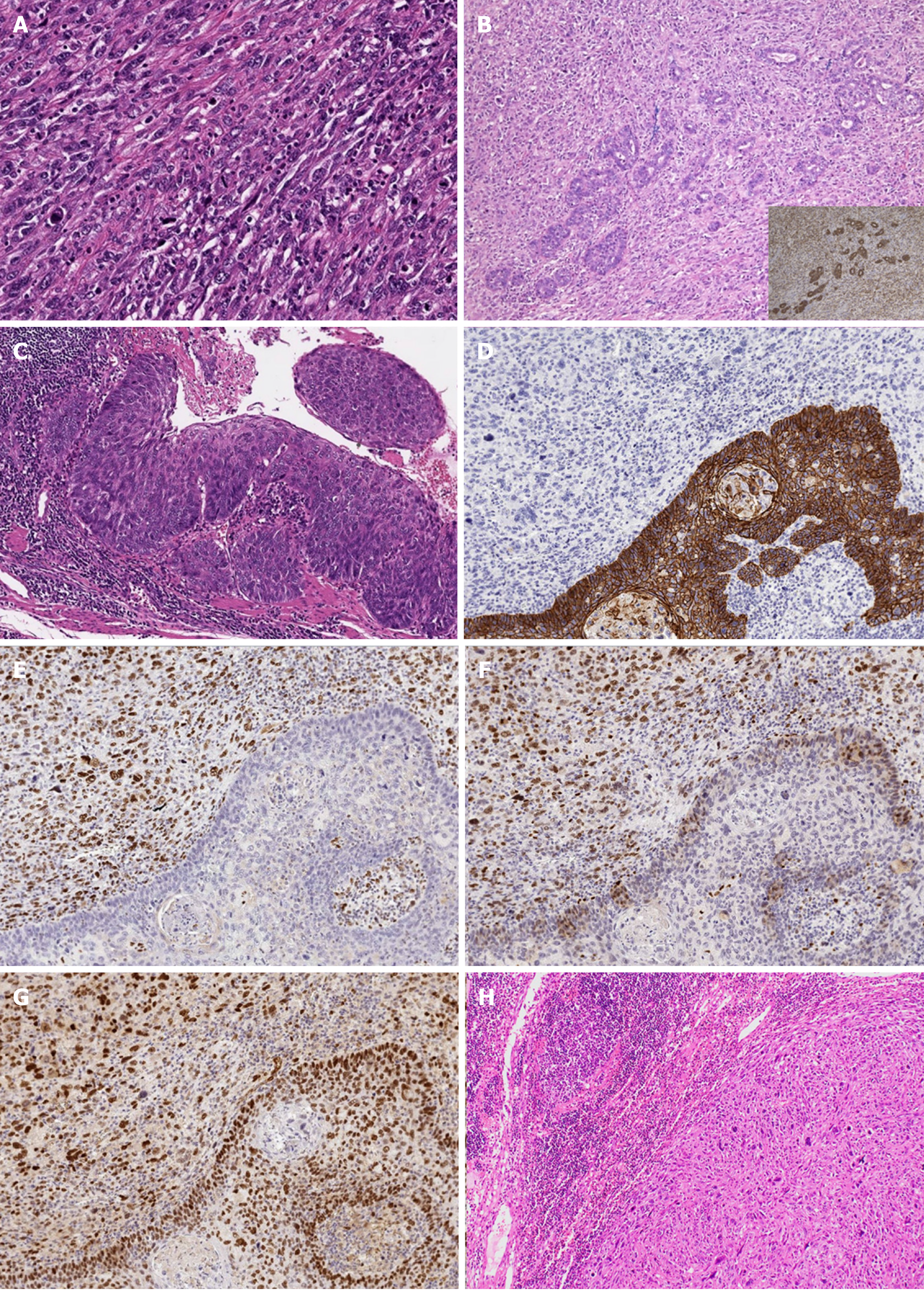
Figure 4 Histopathological and immunohistochemical findings.
A: Dense proliferation and infiltration of spindle cells mixed with giant cells with large atypical nuclei and polynuclear cells were observed (hematoxylin–eosin staining, × 200); B: Adeno-carcinomatous components were detected in a section near the root (hematoxylin–eosin staining, × 100; inserted figure: immunohistochemical expression of CAM 5.2, × 100); C: Squamous cell carcinoma in situ was found in the mucosa of the basal part of the tumor (hematoxylin–eosin staining, × 100); D: E-cadherin was not detected in spindle cells but was detected in the regions of squamous cell carcinoma (× 100); E-G: Epithelial-mesenchymal transition-related markers, zinc finger E-box-binding homeobox 1 (E, × 100), TWIST (F, × 100), and snail family transcriptional repressor 2 (G, × 100) were detected in spindle cells; H: Lymph node metastases with sarcomatous components (hematoxylin–eosin staining, × 40).
- Citation: Okamoto H, Kikuchi H, Naganuma H, Kamei T. Multiple carcinosarcomas of the esophagus with adeno-carcinomatous components: A case report. World J Gastroenterol 2020; 26(17): 2111-2118
- URL: https://www.wjgnet.com/1007-9327/full/v26/i17/2111.htm
- DOI: https://dx.doi.org/10.3748/wjg.v26.i17.2111


