Copyright
©The Author(s) 2020.
World J Gastroenterol. Apr 7, 2020; 26(13): 1439-1449
Published online Apr 7, 2020. doi: 10.3748/wjg.v26.i13.1439
Published online Apr 7, 2020. doi: 10.3748/wjg.v26.i13.1439
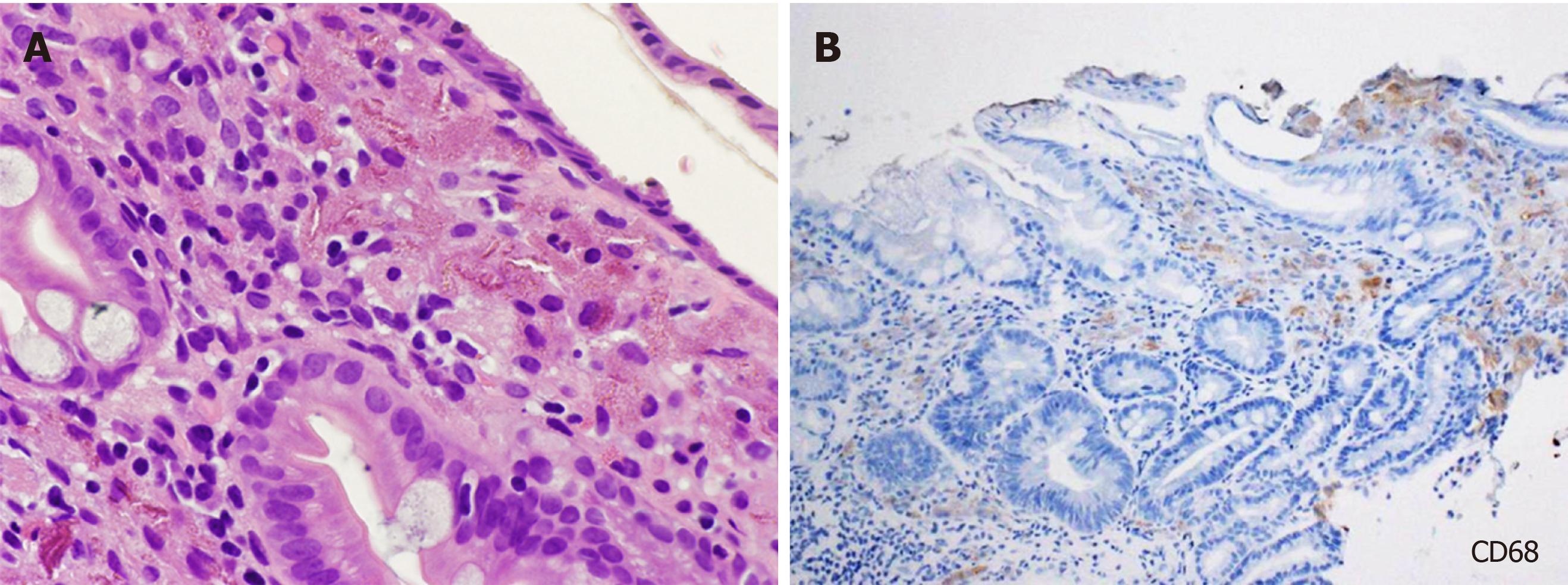
Figure 1 Light microscopy images of gastric lanthanum deposition.
A: Fine, amorphous, eosinophilic material is observed in hematoxylin and eosin staining; B: Deposited material is generally phagocytosed by macrophages.
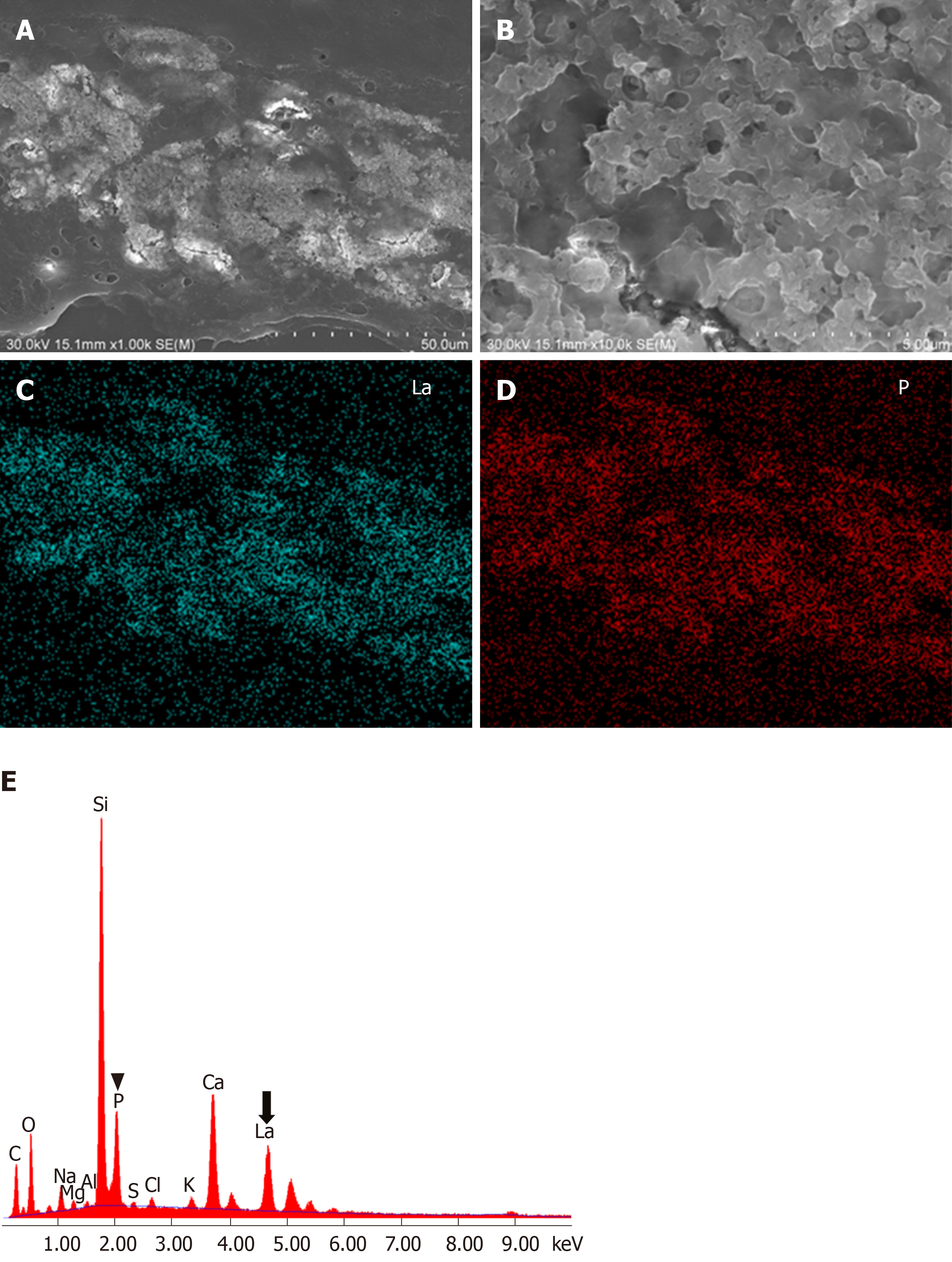
Figure 2 Electron microscopy analysis of gastric lanthanum deposition.
A: In scanning electron microscopy, deposited lanthanum is visible as bright areas; B: Deposited lanthanum is composed of aggregates of particles; C: Elemental mapping with energy dispersive X-ray spectroscopy shows that the distribution of lanthanum; D: Phosphate is identical to that of the bright areas; E: Energy dispersive X-ray spectroscopy shows presence of lanthanum (La) and phosphate elements (P).
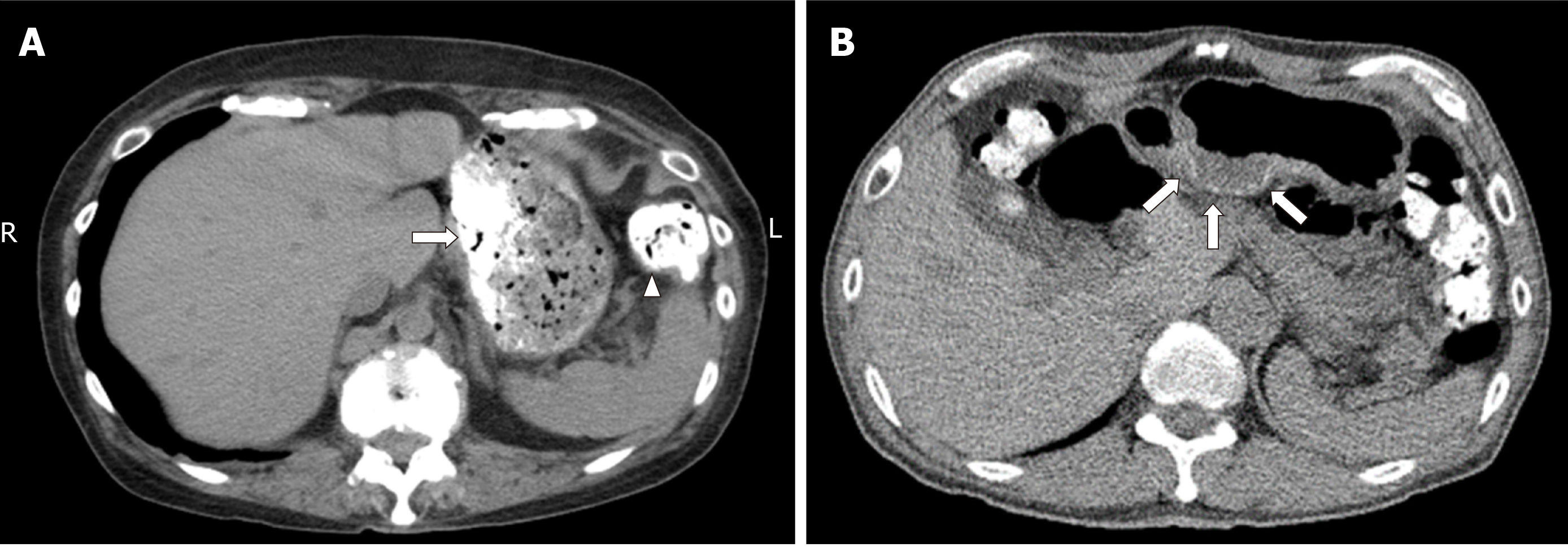
Figure 3 Computed tomography images.
A: Computed tomography scanning shows ingested lanthanum carbonate in the stomach (arrow) and colon (arrowhead) as high-density substances; B: In another patient, deposited lanthanum in the stomach is observed as a high-density layer within the gastric mucosa (arrows).
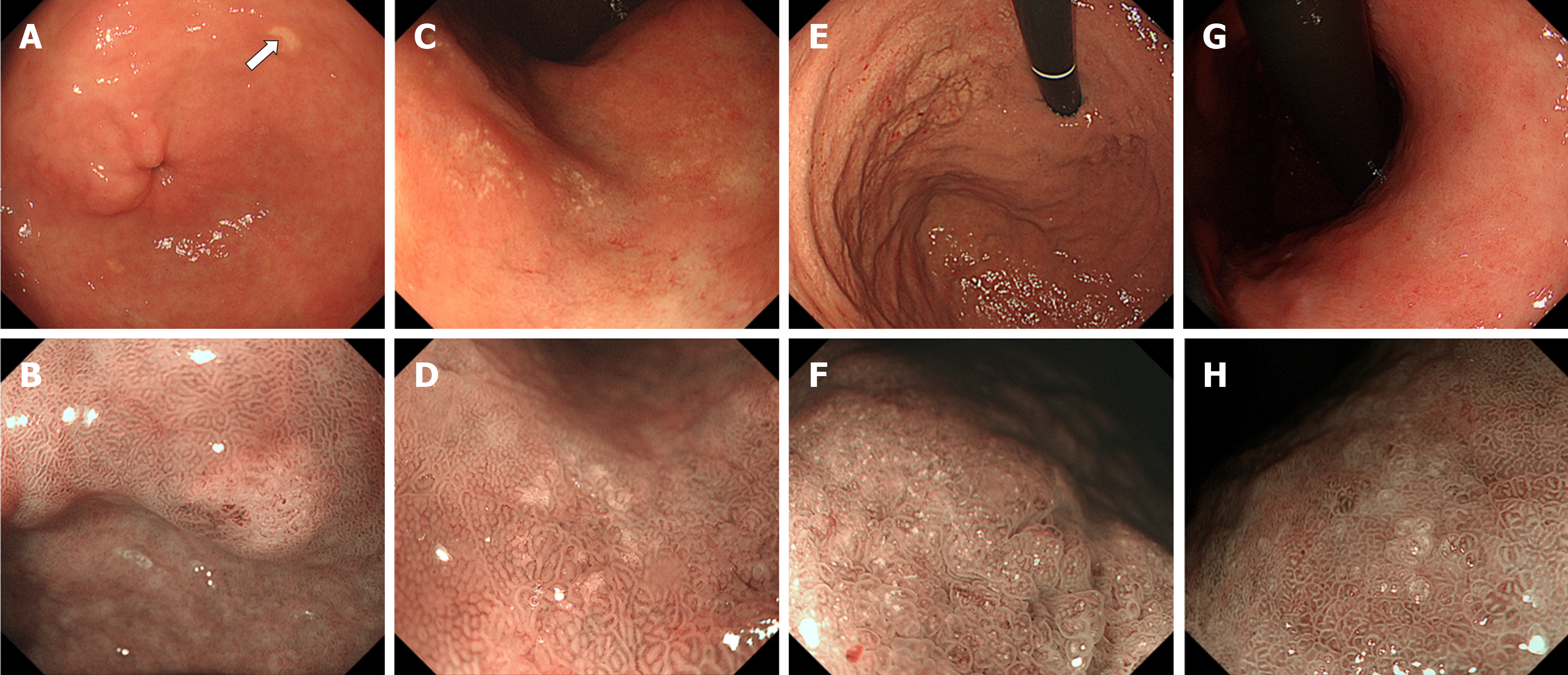
Figure 4 Typical endoscopic features of gastric lanthanum deposition during conventional white-light (upper row) and magnified observation with narrow-band imaging (lower row).
A, B: “Whitish spot” is defined as a whitish lesion ≤ 20 mm in diameter with a uniform white color; C, D: “Annular whitish mucosa” are lesion (s) ≤ 20 mm in diameter with white color in the periphery; E, F: “Diffuse whitish mucosa” appears with a white area > 20 mm in diameter; G, H: “Fine granular whitish deposition” is a tiny or faint whitish lesion (s) ≤ 1 mm in diameter.
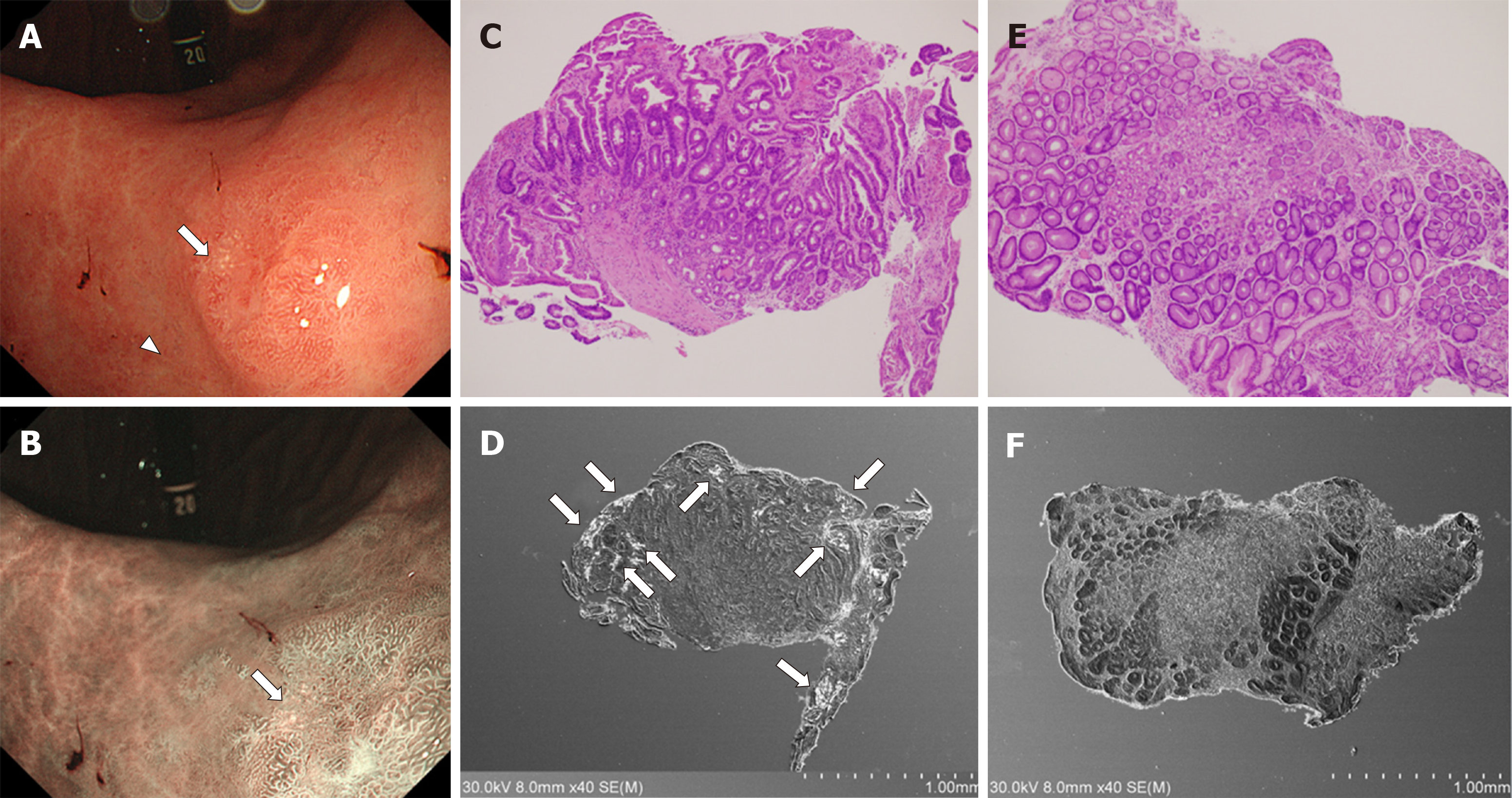
Figure 5 Correlation between intestinal metaplasia and lanthanum deposition in the stomach.
A: Esophagogastroduodenoscopy shows an annular whitish lesion in the gastric antrum (white light observation); B: Esophagogastroduodenoscopy shows an annular whitish lesion in the gastric antrum (narrow-band imaging); C, D: Biopsy sample acquired from a white lesion (A, B, arrows) contains intestinal metaplasia (C) and lanthanum deposition (D); E, F: In contrast, the sample acquired from the surrounding mucosa approximately 5 mm away from the white lesions (A, arrowhead) contains no intestinal metaplasia (E) and lanthanum deposition is subtle (F).
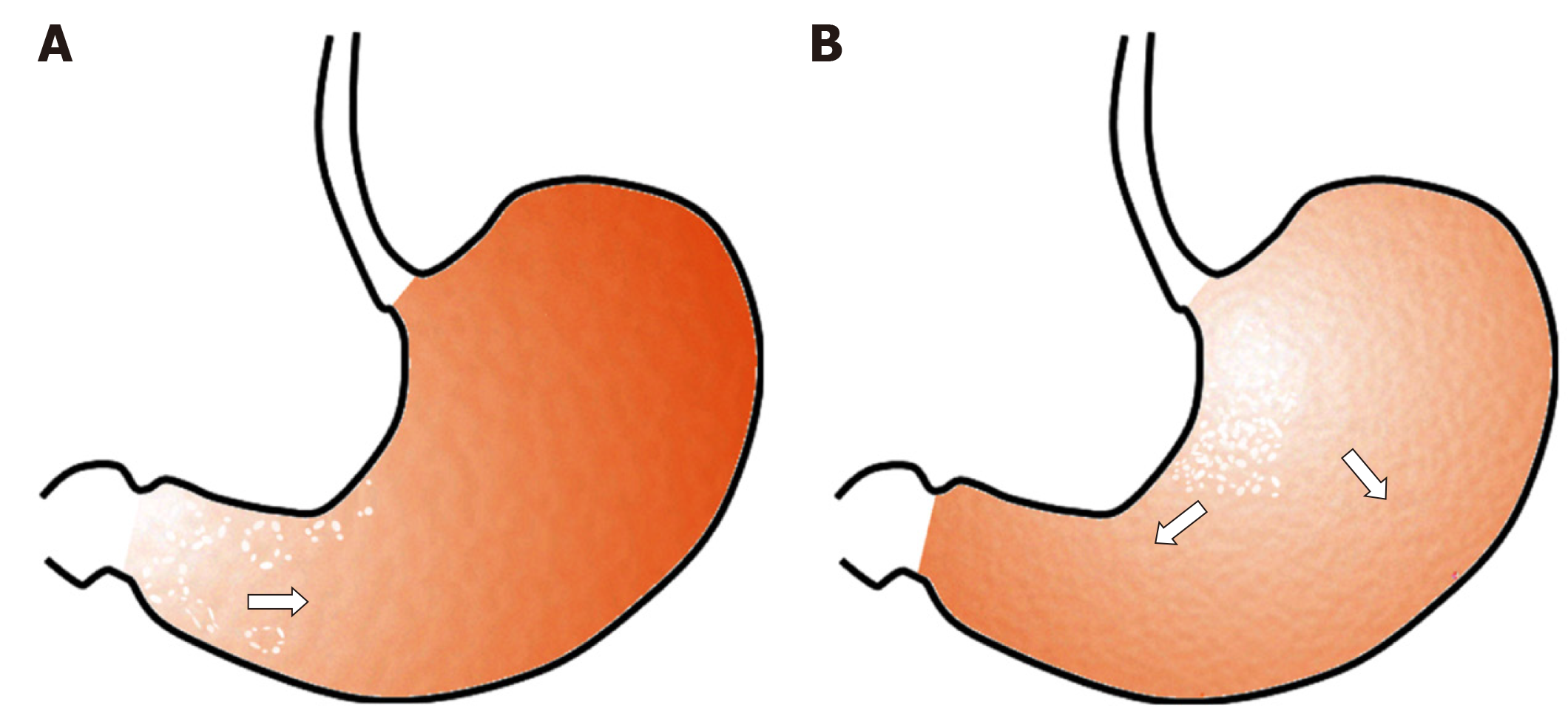
Figure 6 Hypothesis regarding the pattern of lanthanum deposition in the gastric mucosa with or without atrophy.
A: In the atrophic mucosa, particularly in areas with intestinal metaplasia, lanthanum deposition probably presents with annular and/or granular white lesions, predominantly in the gastric antrum and angle. As intestinal metaplasia expands, the size of the areas with lanthanum deposition may increase; B: In the gastric mucosa without atrophy, lanthanum primarily deposits in the lesser curvature and posterior wall of the gastric body and presents diffuse whitish lesions. The area of lanthanum deposition probably expands as time elapses unless the patient stops lanthanum carbonate.
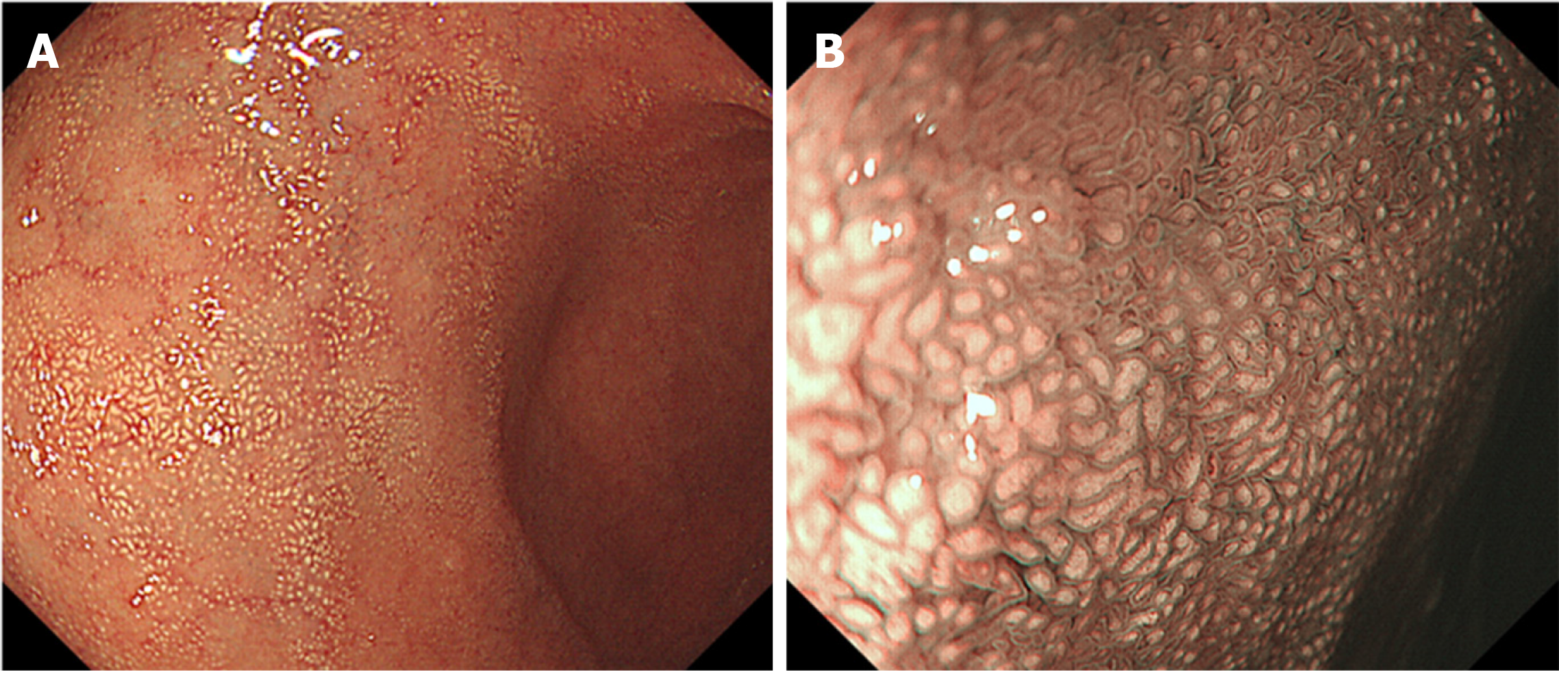
Figure 7 Endoscopic images of the duodenal lanthanum deposition.
A: A lanthanum-related lesion in the duodenum presents with white mucosa; B: Magnified image with narrow-band imaging shows white depositions within the duodenal villi.
- Citation: Iwamuro M, Urata H, Tanaka T, Okada H. Review of the diagnosis of gastrointestinal lanthanum deposition. World J Gastroenterol 2020; 26(13): 1439-1449
- URL: https://www.wjgnet.com/1007-9327/full/v26/i13/1439.htm
- DOI: https://dx.doi.org/10.3748/wjg.v26.i13.1439


