Copyright
©The Author(s) 2020.
World J Gastroenterol. Mar 14, 2020; 26(10): 1080-1087
Published online Mar 14, 2020. doi: 10.3748/wjg.v26.i10.1080
Published online Mar 14, 2020. doi: 10.3748/wjg.v26.i10.1080
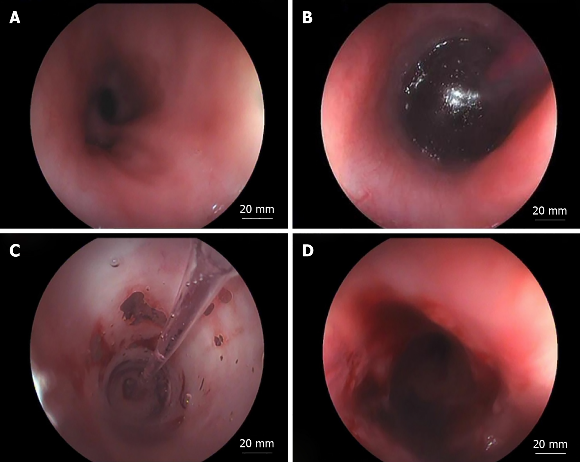
Figure 1 An esophageal stricture in endoscopic view before and after dilation.
A: Lumen tortuosity, deformation, and diverticulisation; B: Mucosal pallor surrounding the inflated balloon; C: Laceration of the mucus observed through the balloon; D: Bleeding and laceration of the esophageal mucus.
- Citation: Dai DL, Zhang CX, Zou YG, Yang QH, Zou Y, Wen FQ. Predictors of outcomes of endoscopic balloon dilatation in strictures after esophageal atresia repair: A retrospective study. World J Gastroenterol 2020; 26(10): 1080-1087
- URL: https://www.wjgnet.com/1007-9327/full/v26/i10/1080.htm
- DOI: https://dx.doi.org/10.3748/wjg.v26.i10.1080


