Copyright
©The Author(s) 2020.
World J Gastroenterol. Mar 14, 2020; 26(10): 1056-1066
Published online Mar 14, 2020. doi: 10.3748/wjg.v26.i10.1056
Published online Mar 14, 2020. doi: 10.3748/wjg.v26.i10.1056
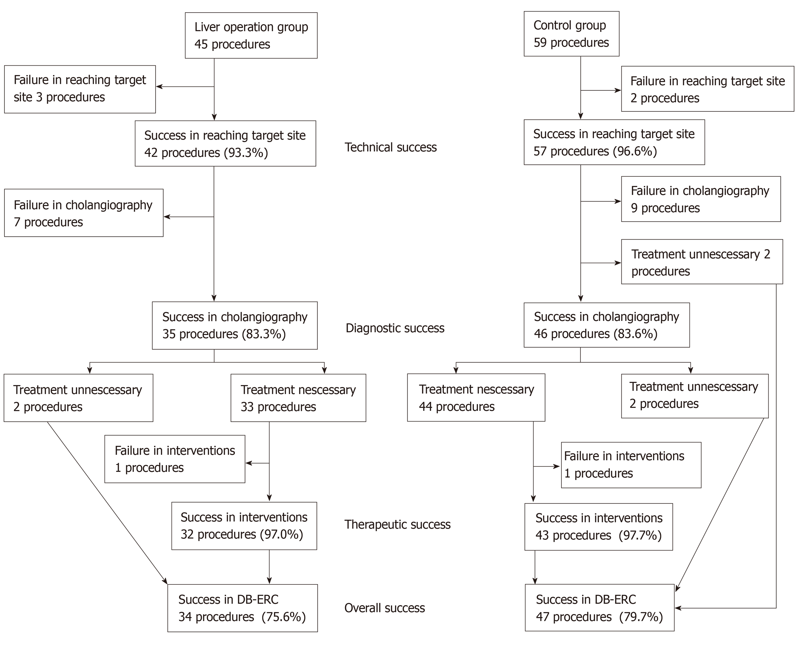
Figure 1 Flowchart of patients’ process.
Technical success: Endoscope reached the hepaticojejunostomy site; Diagnostic success: Successful cholangiography after cannulation to the bile duct; Therapeutic success: Completed interventions in the bile duct; Overall success: Completed diagnosis and interventions among all cases.
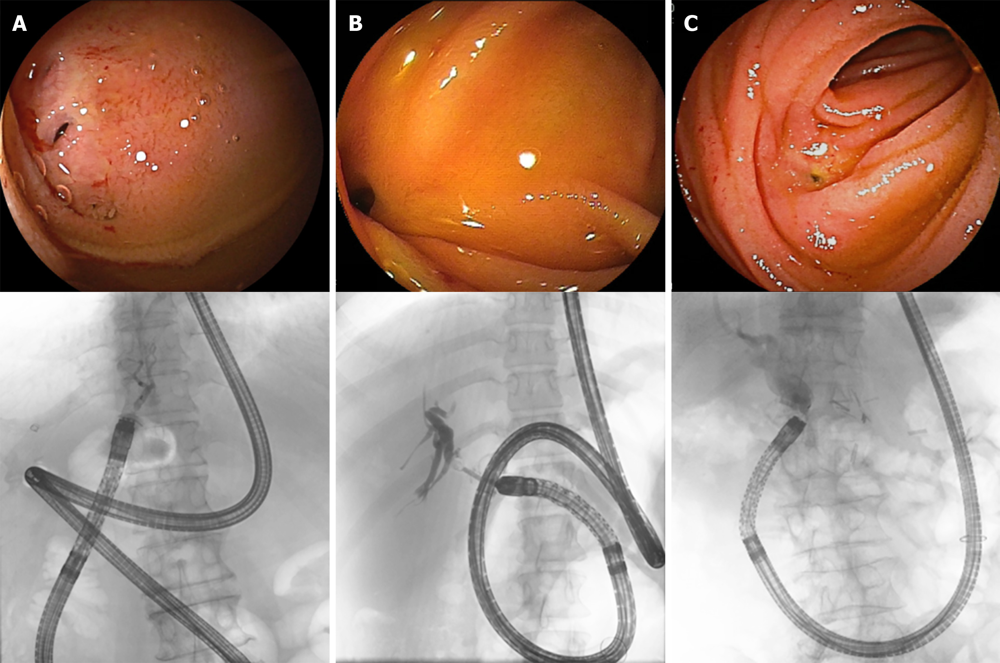
Figure 2 Endoscopic and fluoroscopic images of double-balloon endoscopic retrograde cholangiography.
A: After right hepatic lobectomy; B: After living donor liver transplantation (right lobe graft); C: After pancreatoduodenectomy. These are endoscopic (upper) and fluoroscopic (lower) images of double-balloon endoscopic retrograde cholangiography. Endoscopic image: In A and B, procedures were difficult because the hepaticojejunostomy site was located near the edge of the visual field and close to the endoscope. In C, the hepaticojejunostomy site was located at the center of the visual field, and the distance from the endoscope was appropriate. Fluoroscopic image: In A and B, the endoscope bowed to the side of the removed liver, but no such bowing is observed in C.
- Citation: Nishio R, Kawashima H, Nakamura M, Ohno E, Ishikawa T, Yamamura T, Maeda K, Sawada T, Tanaka H, Sakai D, Miyahara R, Ishigami M, Hirooka Y, Fujishiro M. Double-balloon endoscopic retrograde cholangiopancreatography for patients who underwent liver operation: A retrospective study. World J Gastroenterol 2020; 26(10): 1056-1066
- URL: https://www.wjgnet.com/1007-9327/full/v26/i10/1056.htm
- DOI: https://dx.doi.org/10.3748/wjg.v26.i10.1056


