Copyright
©The Author(s) 2020.
World J Gastroenterol. Jan 7, 2020; 26(1): 35-54
Published online Jan 7, 2020. doi: 10.3748/wjg.v26.i1.35
Published online Jan 7, 2020. doi: 10.3748/wjg.v26.i1.35
Figure 1 Abdominal paracentesis drainage in rats with severe acute pancreatitis.
A: Abdominal paracentesis drainage device; B: Sodium taurocholate (5%) was injected into the biliopancreatic duct using a micro infusion pump; C: Ascitic fluid drained by abdominal paracentesis drainage.
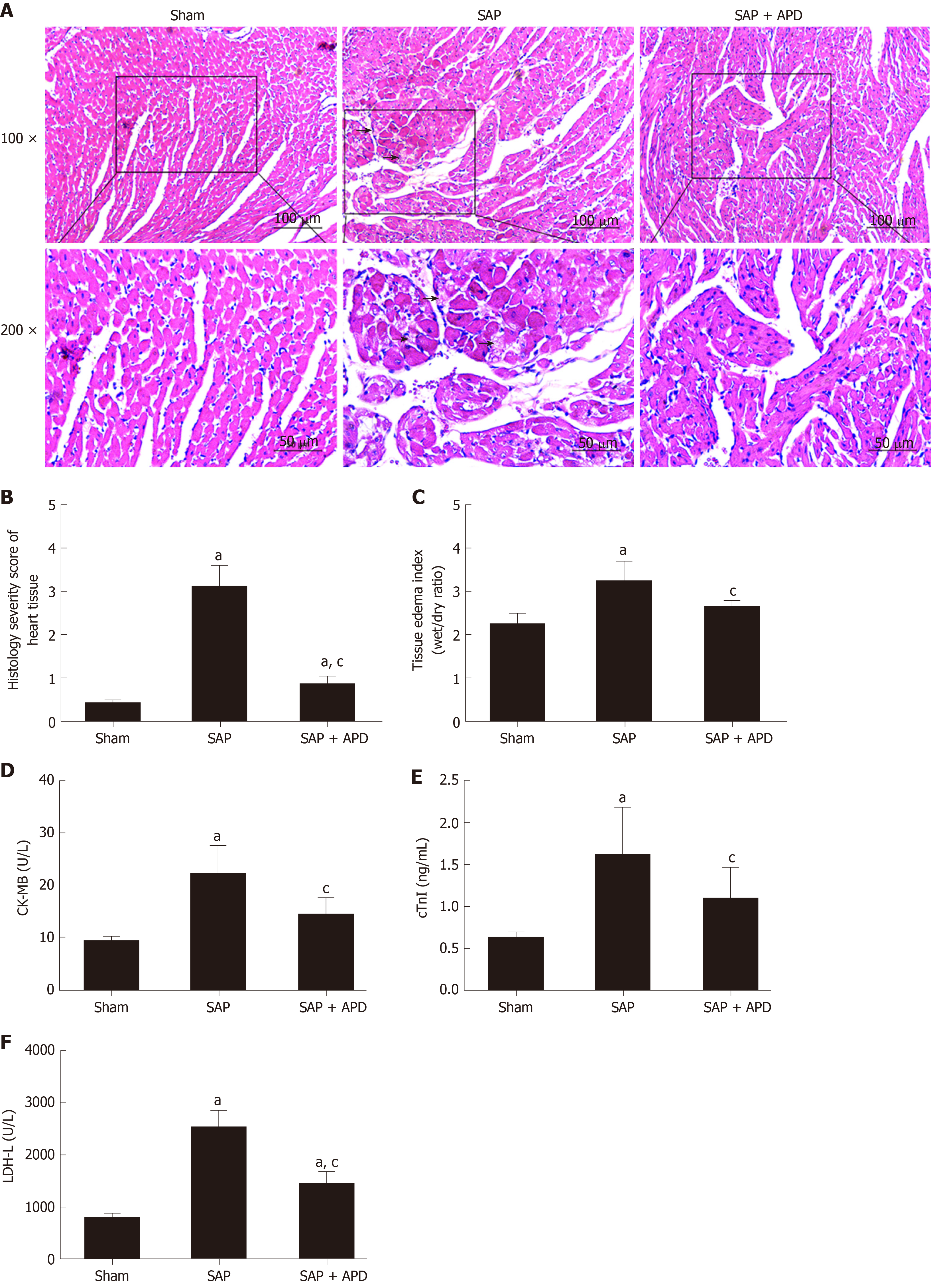
Figure 2 Effects of abdominal paracentesis drainage on cardiac histopathology, tissue edema index and cardiac-related enzymes 24 h after severe acute pancreatitis induction.
A: Representative micrographs of hematoxylin-eosin stained sections of rat heart tissue from different groups. The arrow shows disruptive myocardial fibers. Images were taken under 100 × and 200 × magnification; B: Histology severity score of the heart; C: Tissue edema index of the heart; D-F: The serum levels of Creatine Kinase Isoenzyme MB, Cardiac troponin-I and Lactic Dehydrogenase-L, respectively. Data indicate the mean ± standard deviation obtained from six animals in each group (C-E). aP < 0.05 vs sham group; cP < 0.05 vs severe acute pancreatitis group. SAP: Severe acute pancreatitis; APD: Abdominal paracentesis drainage.
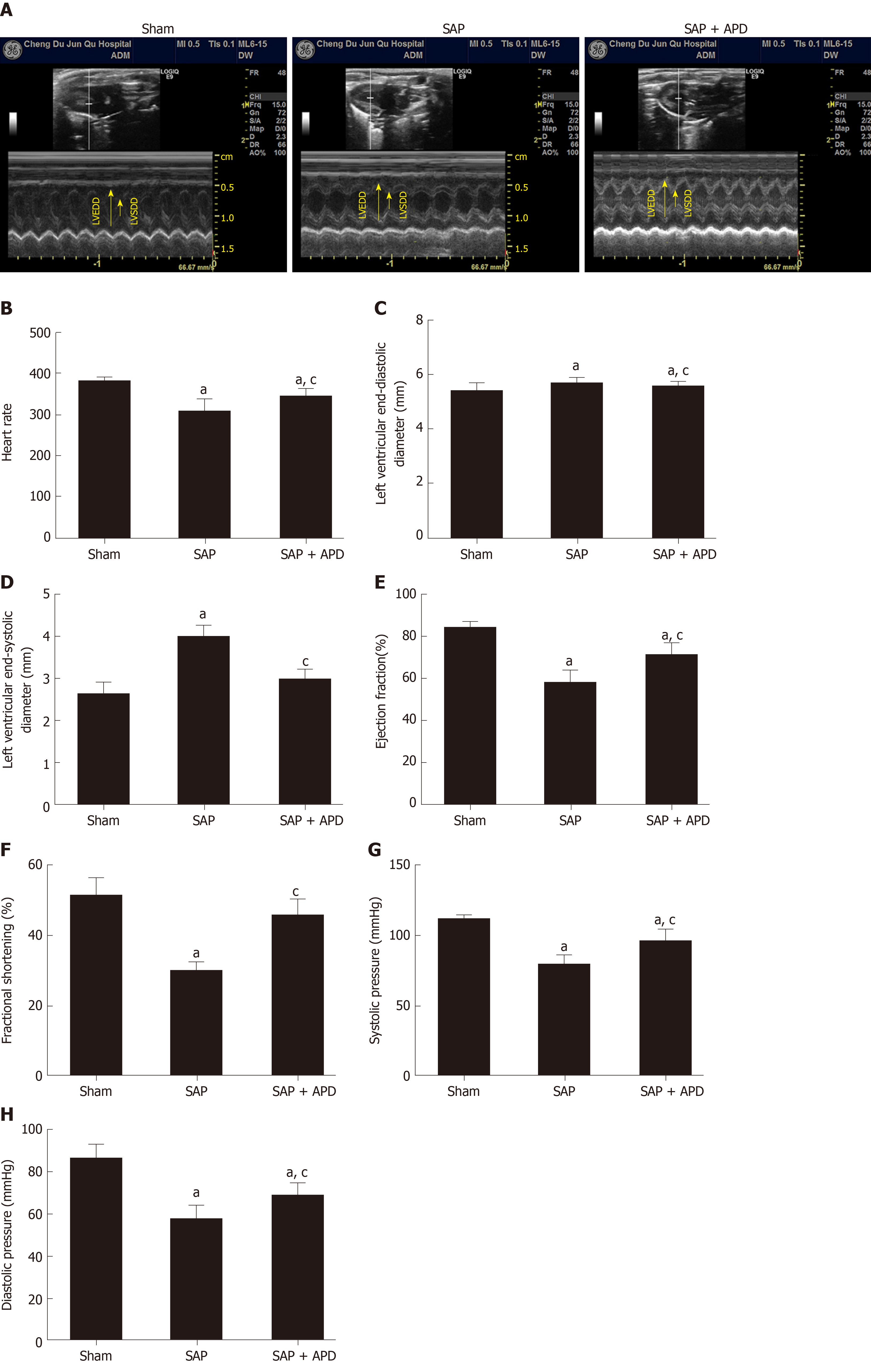
Figure 3 Effect of abdominal paracentesis drainage on echocardiographic and hemodynamic properties.
A: Representative M-mode images in groups; B: Heart rate; C: Left ventricular end-diastolic diameter (mm); D: Left ventricular end-systolic diameter (mm); E: Ejection fraction (%); F: Fractional shortening (%); G: Systolic pressure (mmHg); H: Diastolic pressure (mmHg). Data indicate the mean ± standard deviation obtained from six animals in each group. aP < 0.05 vs sham group; cP < 0.05 vs severe acute pancreatitis group. SAP: Severe acute pancreatitis; APD: Abdominal paracentesis drainage.
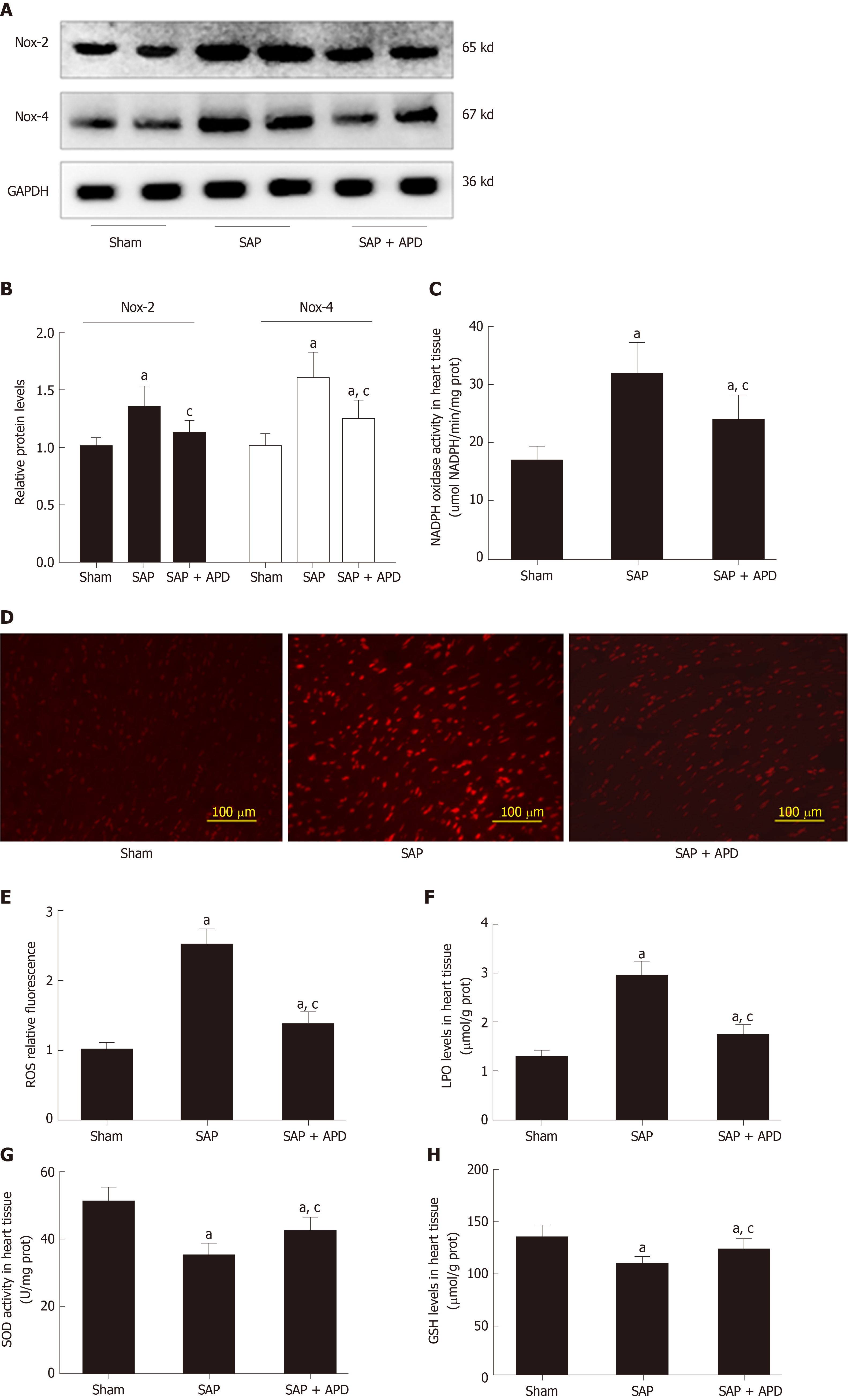
Figure 4 Effect of abdominal paracentesis drainage on the protein expression and activity of nicotinamide adenine dinucleotide phosphate oxidase, reactive oxygen species production and oxidative indexes.
A: Immunoblot of nicotinamide adenine dinucleotide phosphate oxidase-2 (NOX2) and NOX4 protein expression from heart samples; B: Densitometry analysis of NOX2 and NOX4; C: NOX activity was measured by the colorimetric method in heart tissues; D: Dihydroethidium staining in frozen sections by fluorescent microscopy (100 × magnification); E: The fluorescence values are expressed as the ratio to the levels in the sham group; F-H: The levels of lipid peroxidation, the activity of superoxide dismutase and reduced glutathione in heart tissues, respectively. Data are representative of at least three independent experiments (A-B). Data indicate the mean ± standard deviation obtained from six animals in each group (C-H). aP < 0.05 vs sham group; cP < 0.05 vs severe acute pancreatitis group. NOX2: Nicotinamide adenine dinucleotide phosphate oxidase-2; NOX4: Nicotinamide adenine dinucleotide phosphate oxidase-4; GAPDH: Glyceraldehyde-3-phosphate dehydrogenase; SAP: Severe acute pancreatitis; APD: Abdominal paracentesis drainage; ROS: Reactive oxygen species; LPO: Lipid peroxidation; SOD: Superoxide dismutase; GSH: Reduced glutathione.
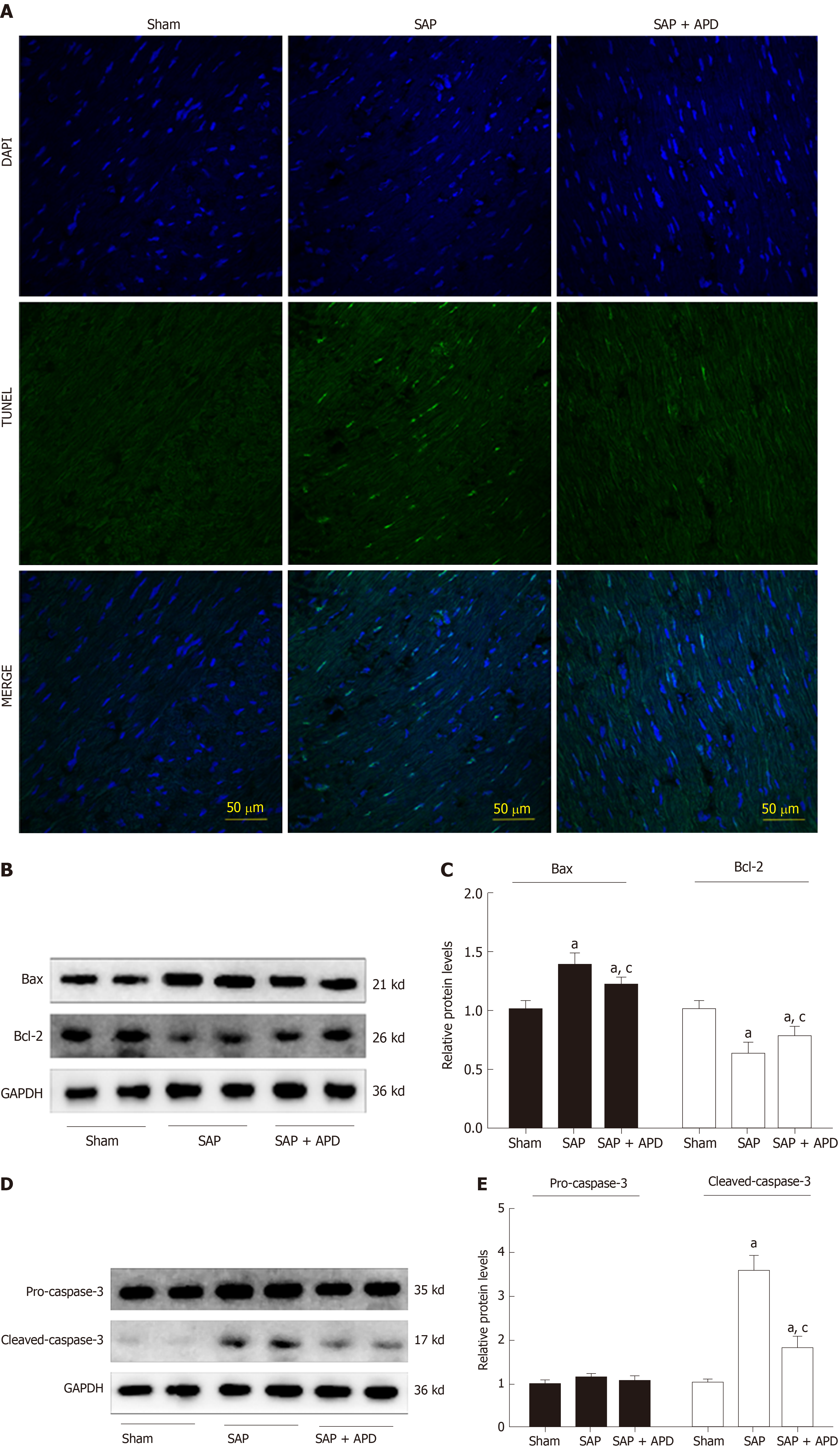
Figure 5 Abdominal paracentesis drainage attenuates myocardial cell apoptosis and expression of apoptosis associated protein in myocardium.
A: Representative images (400 × magnification) of terminal deoxynucleotidyl transferase-mediated dUTP nick end labeling assay. Cells with green staining in the nuclei were determined to be apoptotic cells; B: Immunoblot of Bax and Bcl-2 protein expression from heart samples; C: Densitometry analysis of Bax and Bcl-2. Data are representative of at least three independent experiments; D: Immunoblot of pro-caspase-3 and cleaved-caspase-3 protein expression from heart samples; E: Densitometry analysis of pro-caspase-3 and cleaved-caspase-3. Data are representative of at least three independent experiments. aP < 0.05 vs sham group; cP < 0.05 vs severe acute pancreatitis group. TUNEL: Terminal deoxynucleotidyl transferase-mediated dUTP nick end labeling; Bax: Bcl-2 related X protein; Bcl-2: B cell lymphoma/leukemia-2 gene; GAPDH: Glyceraldehyde-3-phosphate dehydrogenase; SAP: Severe acute pancreatitis; APD: Abdominal paracentesis drainage.
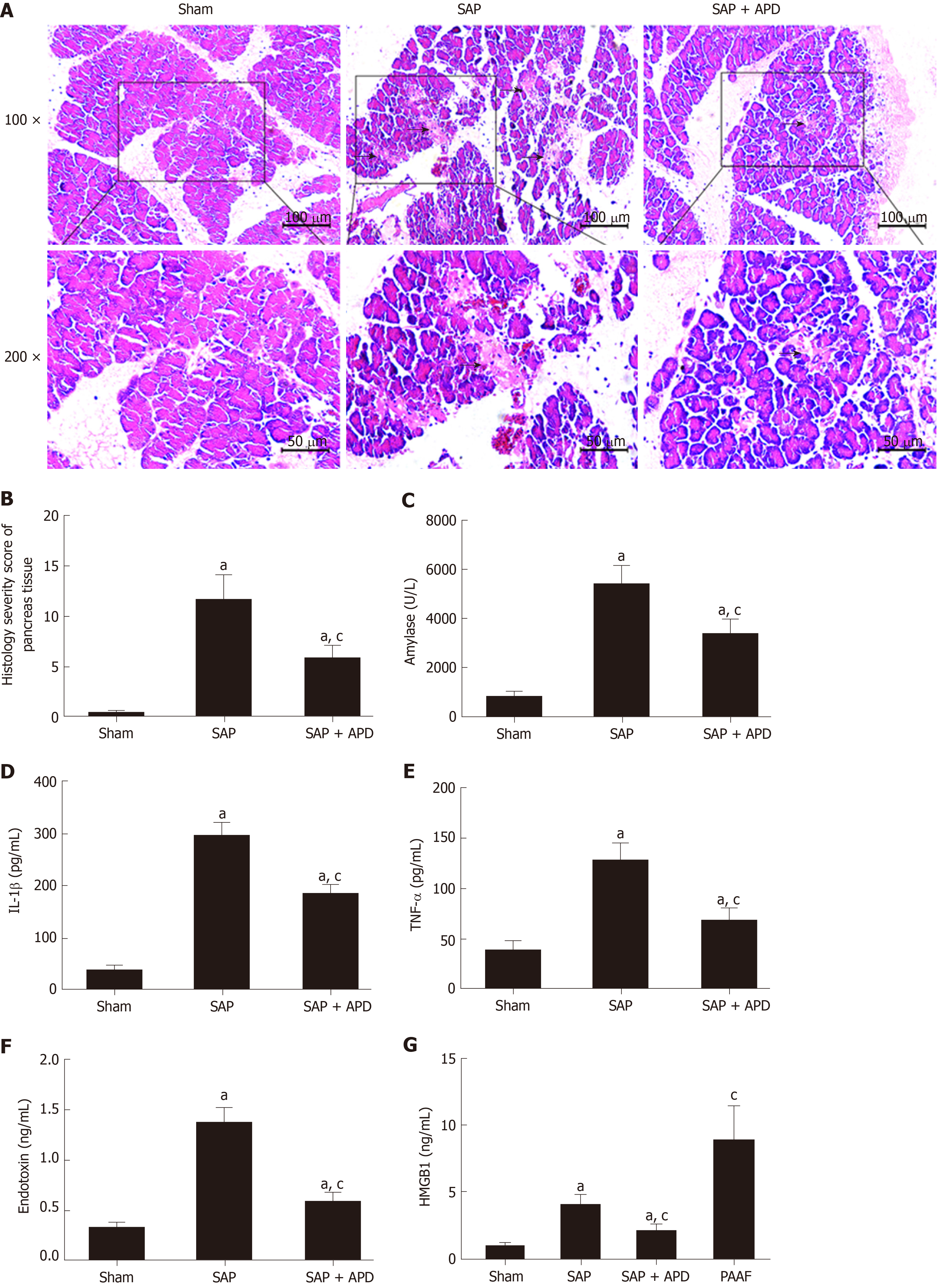
Figure 6 Effects of abdominal paracentesis drainage on pancreatic histopathology and proinflammatory cytokines.
A: Representative micrographs of hematoxylin-eosin stained sections of rat pancreatic tissue from different groups. Images were taken under 100 × and 200 × magnification. The arrow indicates necrotic pancreatic tissue; B: Histology severity score of pancreas; C: Amylase; D: Interleukine-1 beta; E: Tumor necrosis factor alpha; F: Endotoxin; G: High mobility group box 1. Data indicate the mean ± standard deviation obtained from six animals in each group (C-G). aP < 0.05 vs sham group; cP < 0.05 vs severe acute pancreatitis group. IL-1β: Interleukine-1 beta; TNF-α: Tumor necrosis factor alpha; HMGB1: High mobility group box 1; PAAF: Pancreatitis associated ascitic fluids; SAP: Severe acute pancreatitis; APD: Abdominal paracentesis drainage.
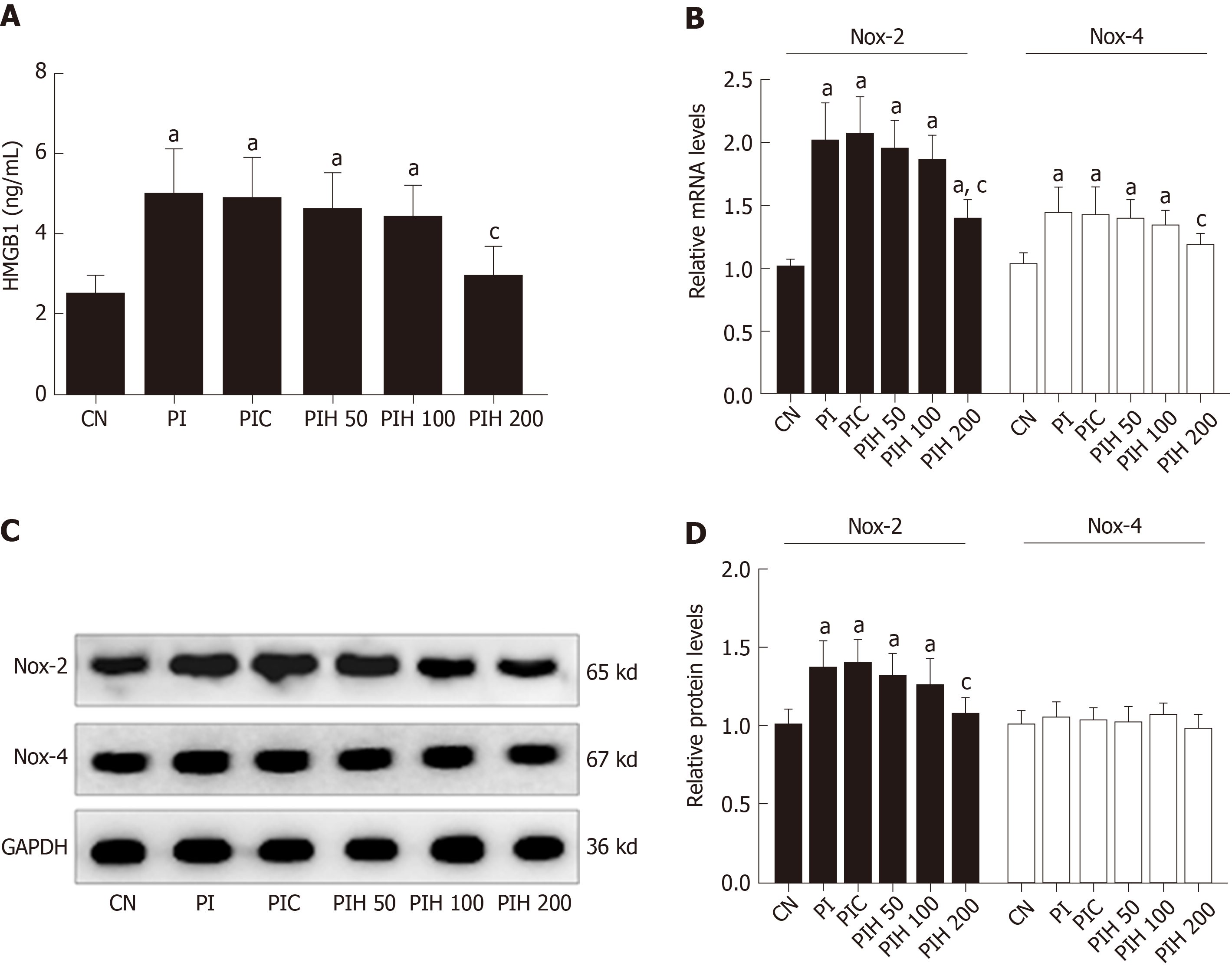
Figure 7 Effects of pancreatitis associated ascitic fluids intraperitoneal injection with or without anti-high mobility group box 1 neutralizing antibody on cardiac NOX expression in cerulein rats.
A: High mobility group box 1 in the serum; B: Nicotinamide adenine dinucleotide phosphate oxidase-2 (NOX2) and NOX4 mRNA measurement by real-time polymerase chain reaction; C: Immunoblot of NOX2 and NOX4 protein expression from heart samples; D: Densitometry analysis of NOX2 and NOX4. Data indicate the mean ± standard deviation obtained from six animals in each group (A-B). Data are representative of at least three independent experiments (C-D). aP < 0.05 vs controls group; cP < 0.05 vs pancreatitis-associated ascitic fluid injection group. HMGB1: High mobility group box 1; NOX2: Nicotinamide adenine dinucleotide phosphate oxidase-2; NOX4: Nicotinamide adenine dinucleotide phosphate oxidase-4; GAPDH: Glyceraldehyde-3-phosphate dehydrogenase; SAP: Severe acute pancreatitis; APD: Abdominal paracentesis drainage.
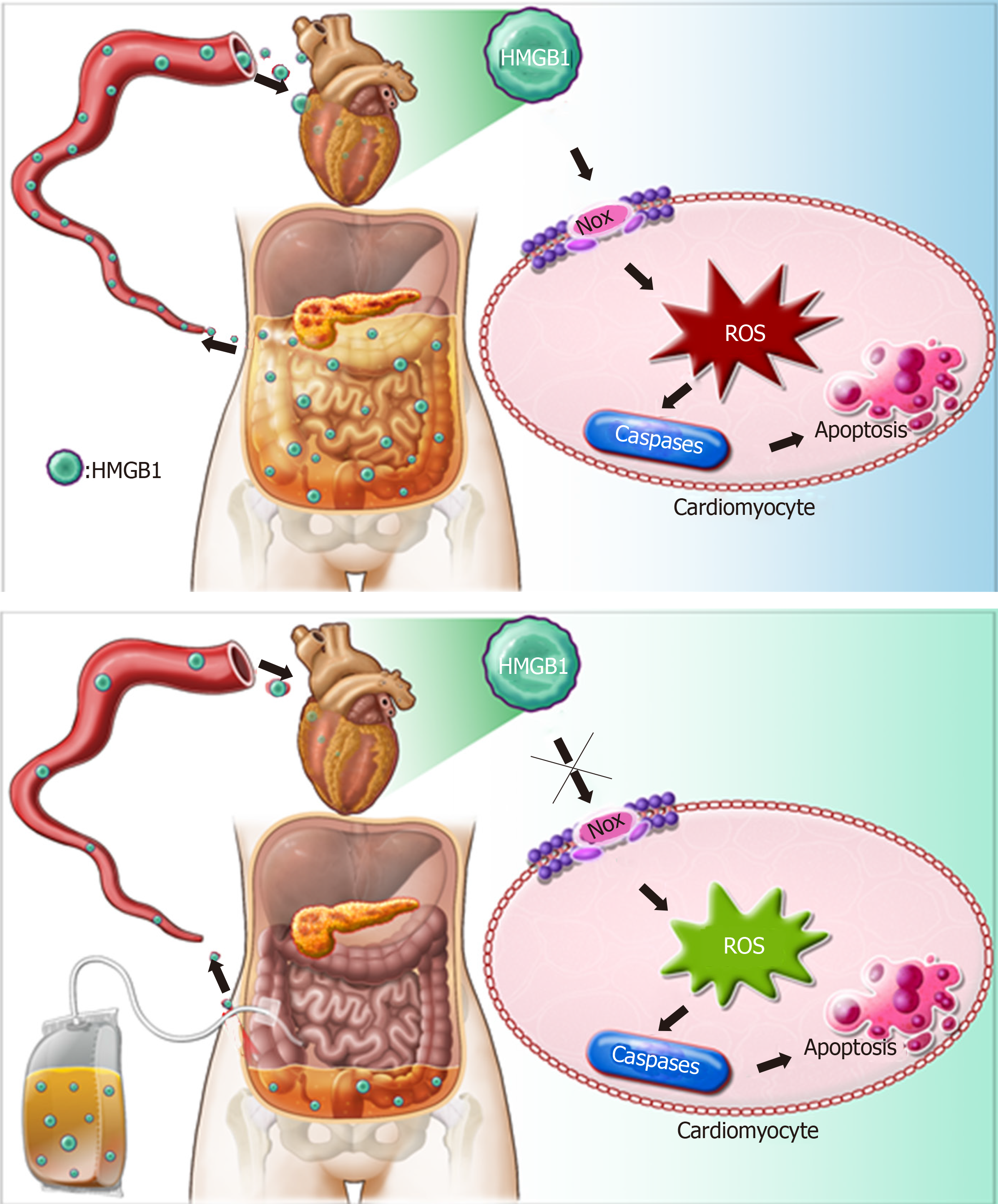
Figure 8 Possible mechanisms responsible for the protective effects of abdominal paracentesis drainage on severe acute pancreatitis associated cardiac injury.
During pancreatitis, high levels of high mobility group box 1 (HMGB1) in the bloodstream can trigger a lethal inflammatory process and participate in the development of remote organ injury. Following abdominal paracentesis drainage treatment, the levels of high mobility group box 1 in the circulation decrease significantly, resulting in inhibition of expression and activity of cardiac nicotinamide adenine dinucleotide phosphate oxidase, thus reactive oxygen species production markedly decreases. These events downregulate expression of caspase-associated proteins and alleviate apoptosis, thereby yielding beneficial effects. HMGB1: High mobility group box 1; APD: Abdominal paracentesis drainage; NOX: Nicotinamide adenine dinucleotide phosphate oxidase; ROS: Reactive oxygen species.
- Citation: Wen Y, Sun HY, Tan Z, Liu RH, Huang SQ, Chen GY, Qi H, Tang LJ. Abdominal paracentesis drainage ameliorates myocardial injury in severe experimental pancreatitis rats through suppressing oxidative stress. World J Gastroenterol 2020; 26(1): 35-54
- URL: https://www.wjgnet.com/1007-9327/full/v26/i1/35.htm
- DOI: https://dx.doi.org/10.3748/wjg.v26.i1.35


