Copyright
©The Author(s) 2019.
World J Gastroenterol. Mar 7, 2019; 25(9): 1050-1066
Published online Mar 7, 2019. doi: 10.3748/wjg.v25.i9.1050
Published online Mar 7, 2019. doi: 10.3748/wjg.v25.i9.1050
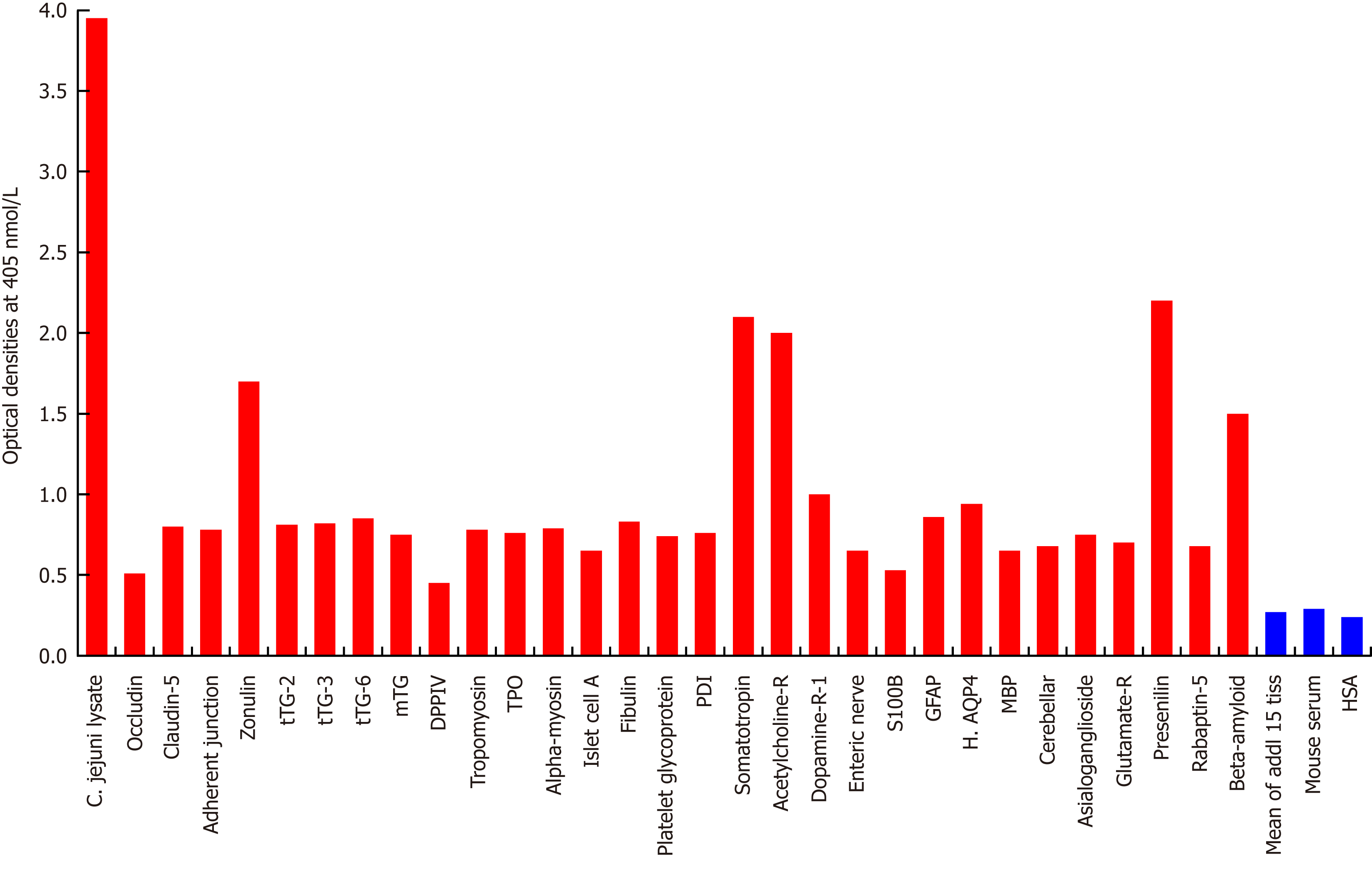
Figure 1 Reaction of mouse monoclonal anti-Campylobacter jejuni with tissue antigens.
Compared to the monoclonal antibody’s reaction with Campylobacter jejuni (C. jejuni) lysate as positive control and human serum albumin (HSA) or unimmunized mouse serum as negative control, the reaction of this antibody with 15 tissue antigens is insignificant, with 25 additional antigens is low, with β-amyloid, zonulin, acetylcholine receptor, somatotropin and presenilin ranges from moderate to high, and with C. jejuni lysate is very highly positive. The variation of quadruplicate ODs was less than 10% for all determinations. Comparison of the means of the antibody reactivity to various tissue antigens with the means of controls resulted in insignificant values (P > 0.05) for 15 different tissue antigens and highly significant P values (P < 0.00001) for the others. 0-0.4 OD = insignificant; 0.42-1.0 = low; 1.1-1.6 = moderate; 1.61-2.2 = high; > 2.21 = very high. tTG: Transglutaminase; mTG: Microbial transglutaminase; DPPIV: Dipeptidyl peptidase-4; TPO: Thyroid peroxidase; PDI: Protein disulfide isomerase; GFAP: Glial fibrillary acidic protein; H. AQP4: Human aquaporin-4; MBP: Myelin basic protein; HSA: Human serum albumin.
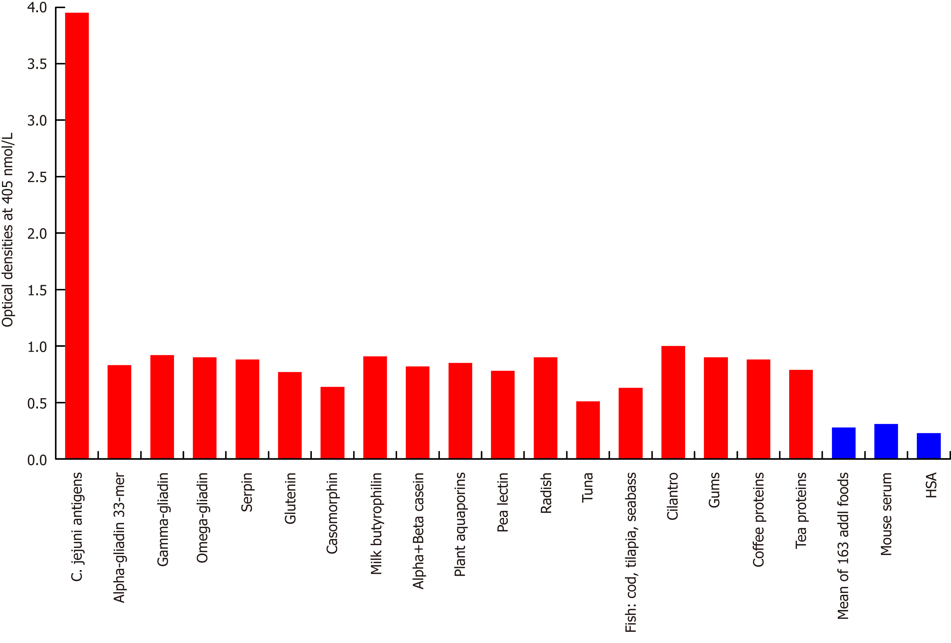
Figure 2 Reaction of mouse monoclonal anti-Campylobacter jejuni with different food antigens.
Compared to the monoclonal antibody’s reaction with Campylobacter jejuni (C. jejuni) antigens as positive control and human serum albumin or unimmunized mouse serum as negative control, the reaction of this antibody with 163 food antigens is insignificant, and with 17 additional antigens is low. The variation of quadruplicate ODs was less than 10% for all determinations. Comparison of the means of the antibody reactivity to various food antigens with the means of controls resulted in insignificant values (P > 0.05) for 163 different food antigens and highly significant P values (P < 0.00001) for the other 17 antigens. HSA: Human serum albumin.
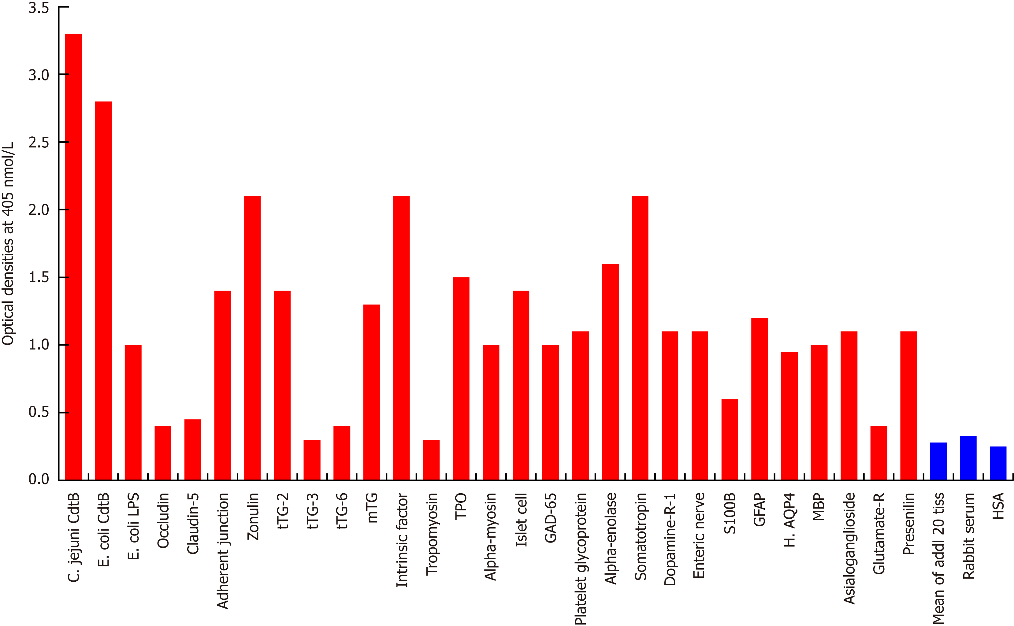
Figure 3 Reaction of affinity-purified rabbit anti-Campylobacter jejuni cytolethal distending toxin with different tissue antigens.
Compared to the reaction with Campylobacter jejuni (C. jejuni) cytolethal distending toxin (Cdt) as positive control and human serum albumin (HSA) or unimmunized rabbit serum as negative control, the reaction of this antibody with 20 tissue antigens is insignificant, with an additional 5 tissue antigens slightly higher than the mean of the 20 but still insignificant; with Escherichia coli (E. coli) lipopolysaccharide (LPS), α-myosin, claudin-5 and 8 different neuronal or associated antigens (enteric nerve, S100B, AQP4, myelin basic protein, asialoganglioside GM1, presenelin and glutamic acid decarboxylase-65) is low, with adherent junction, transglutaminase-2, microbial transglutaminase, thyroid peroxidase, islet cell, a-enolase, glial fibrillary acidic protein, dopamine-R1, and platelet glycoprotein is moderate, with somatotropin, intrinsic factor and zonulin is high, and with E. coli Cdt is very highly positive. The variation of quadruplicate ODs was less than 10% for all determinations. Comparison of the means of the antibody reactivity to various food antigens with the means of controls resulted in insignificant values (P > 0.05) for 25 different tissue antigens and highly significant P values (P < 0.00001) for the other antigens. E. coli: Escherichia coli; Cdt: Cytolethal distending toxin; LPS: Lipopolysaccharide; tTG: Transglutaminase; mTG: Microbial transglutaminase; DPPIV: Dipeptidyl peptidase-4; TPO: Thyroid peroxidase; GAD-65: Glutamic acid decarboxylase-65; GFAP: Glial fibrillary acidic protein; H. AQP4: Human aquaporin-4; MBP: Myelin basic protein; Glutamate-R: Glutamate receptor; HSA: Human serum albumin.
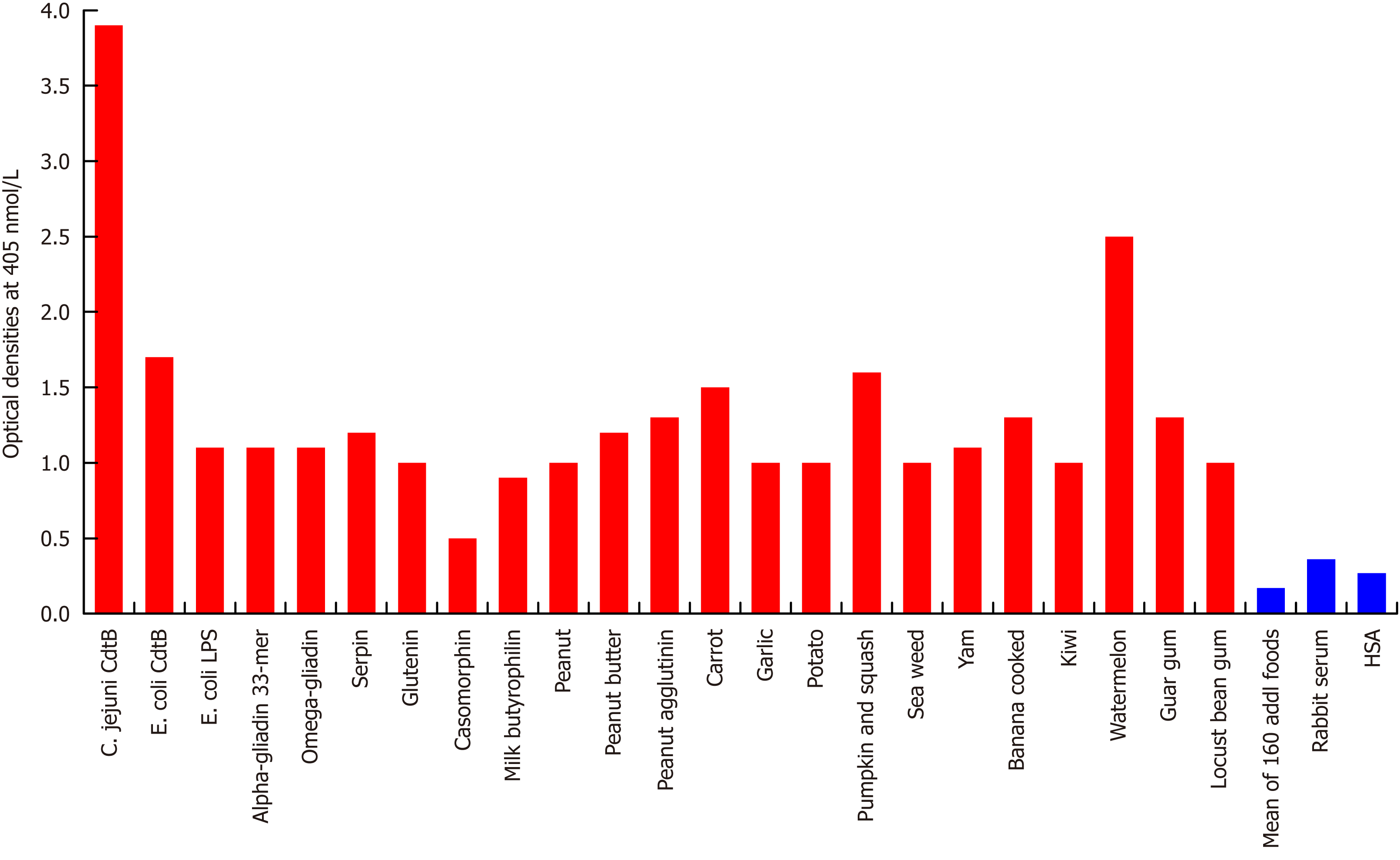
Figure 4 Reaction of affinity-purified rabbit anti-Campylobacter jejuni cytolethal distending toxin with different food antigens.
Compared to the reaction with Campylobacter jejuni (C. jejuni) antigens as positive control and HSA or unimmunized rabbit serum as negative control, the reaction of this antibody with 160 out of 180 food antigens is insignificant, and with 20 additional antigens ranges from low to very high. The variation of quadruplicate ODs was less than 10% for all determinations. Comparison of the means of the antibody reactivity to various food antigens with the means of controls resulted in insignificant values (P > 0.05) for 160 different food antigens and highly significant P values (P < 0.00001) for the other 20 antigens. E. coli: Escherichia coli; Cdt: Cytolethal distending toxin; LPS: Lipopolysaccharide; HSA: Human serum albumin.
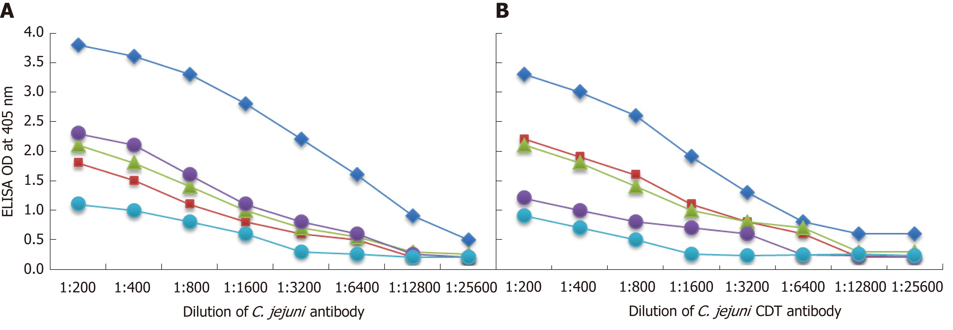
Figure 5 Demonstration of analytical specificity by dilution study.
A: Reaction of various dilutions of mouse monoclonal anti-Campylobacter jejuni (C. jejuni) bacteria antibody with the same concentrations of C. jejuni = blue rhombus, zonulin = red square, somatotropin = green triangle, presenilin = purple circle, and ω-gliadin = blue circle. B: Reaction of various dilutions of mouse monoclonal anti-C. jejuni bacterial cytolethal distending toxin (Cdt) antibody with the same concentrations of C. jejuni Cdt = blue rhombus, zonulin = red square, somatotropin = green triangle, presenilin = purple circle, and ω-gliadin = blue circle. CDT: Cytolethal distending toxin; C. jejuni: Campylobacter jejuni.
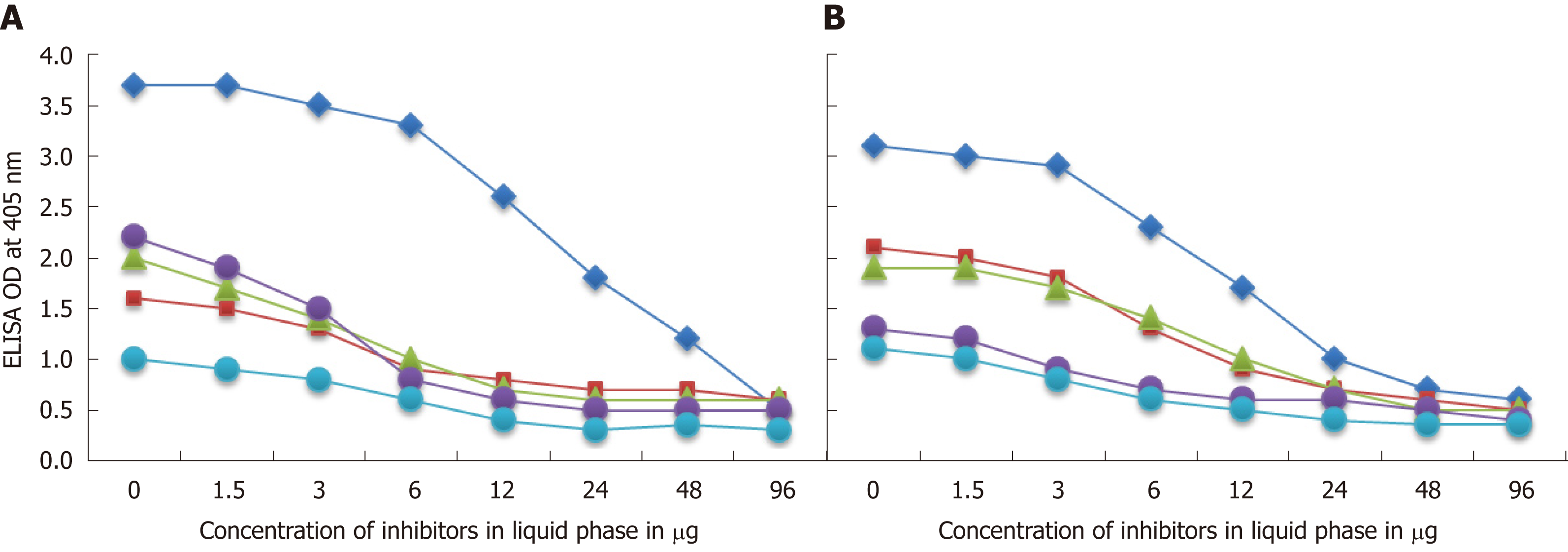
Figure 6 Demonstration of analytical specificity by inhibition study.
A: Inhibition of the reaction of mouse monoclonal anti-Campylobacter jejuni (C. jejuni) antibody binding to C. jejuni antigens = blue rhombus, zonulin = red square, somatotropin = green triangle, presenilin = purple circle, and ω-gliadin = blue circle with different concentrations of the same antigen in liquid phase. B: Inhibition of the reaction of affinity-purified rabbit anti-C. jejuni cytolethal distending toxin antibody binding to C. jejuni cytolethal distending toxin = blue rhombus, zonulin = red square, somatotropin = green triangle, presenilin = purple circle, and ω-gliadin = blue circle with different concentrations of the same antigen in liquid phase.
- Citation: Vojdani A, Vojdani E. Reaction of antibodies to Campylobacter jejuni and cytolethal distending toxin B with tissues and food antigens. World J Gastroenterol 2019; 25(9): 1050-1066
- URL: https://www.wjgnet.com/1007-9327/full/v25/i9/1050.htm
- DOI: https://dx.doi.org/10.3748/wjg.v25.i9.1050


