Copyright
©The Author(s) 2019.
World J Gastroenterol. Nov 28, 2019; 25(44): 6483-6494
Published online Nov 28, 2019. doi: 10.3748/wjg.v25.i44.6483
Published online Nov 28, 2019. doi: 10.3748/wjg.v25.i44.6483
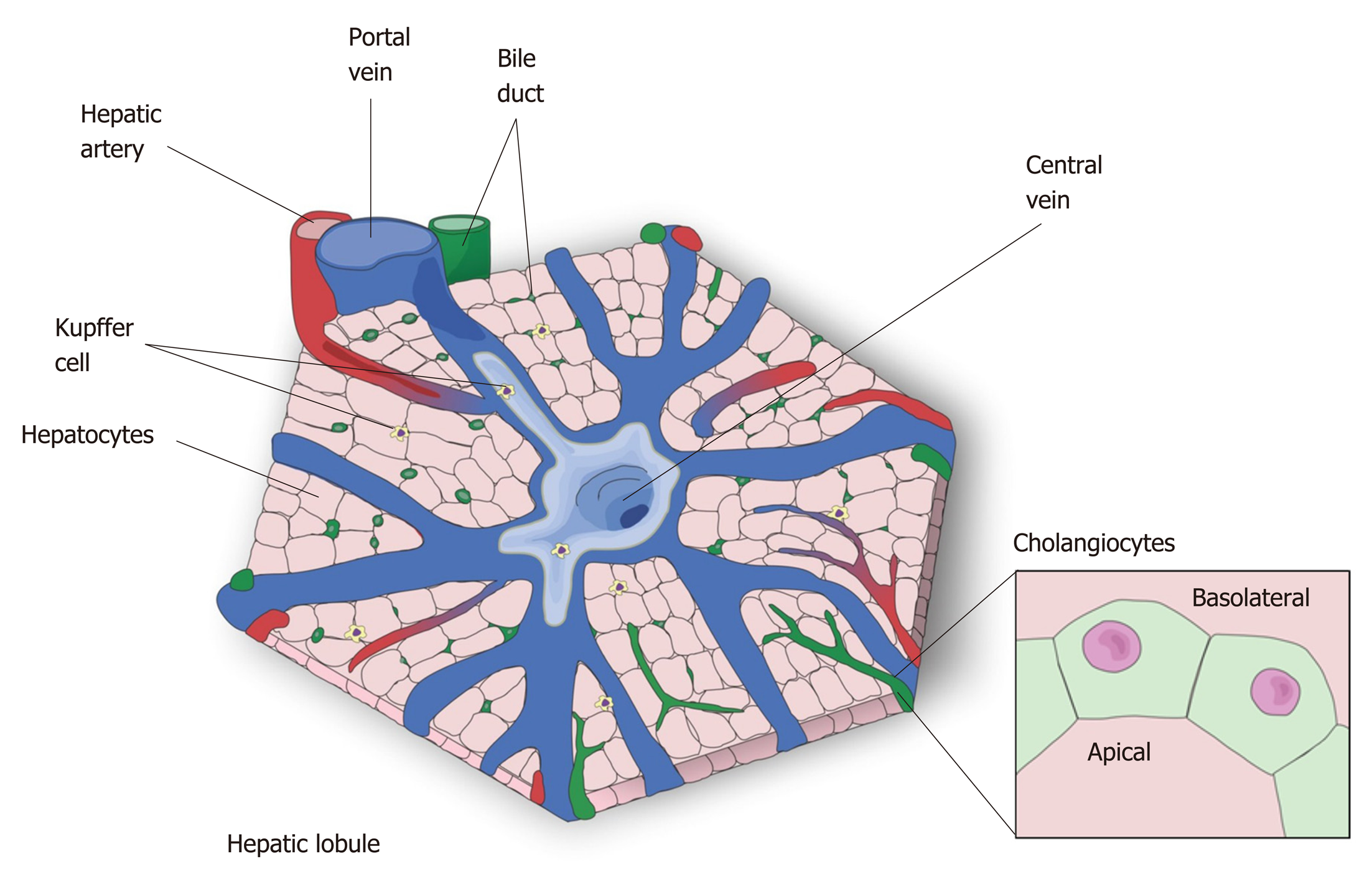
Figure 1 The hepatic lobule-the microscopic functional structure of the liver.
The hepatocytes are arranged in cordon, connecting the central vein to the portal triad, which is formed by a hepatic artery, a portal vein and a bile duct. The other represented cell types are: Kupffer cell, resident macrophages responsible for the immunologic response in the liver, and cholangiocytes which form the bile duct that transport bile to the gallbladder.
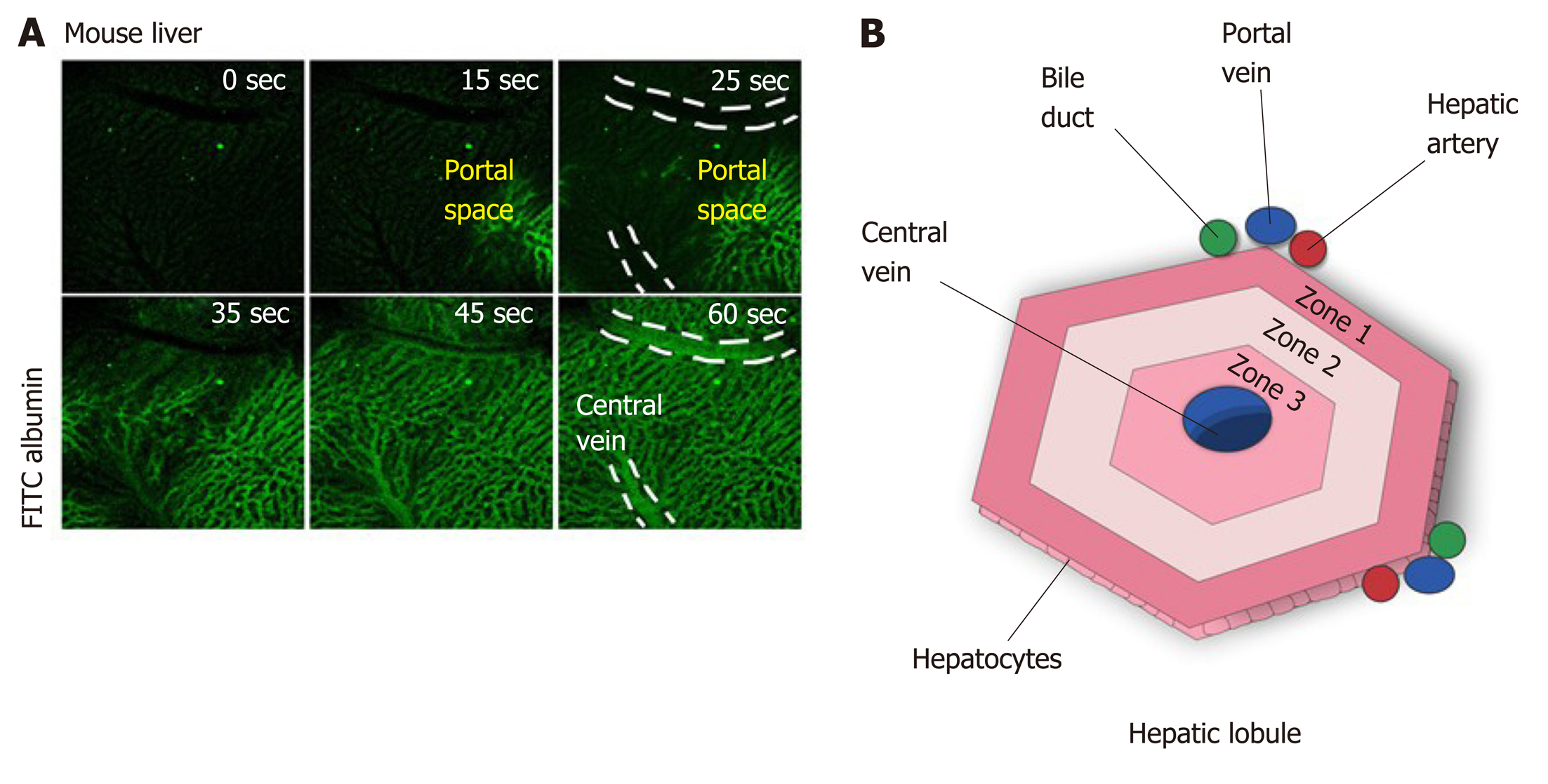
Figure 2 The microenvironment in the hepatic lobules.
A: Perfusion with fluorescein isothiocyanate albumin shows that the blood flow arrives in the liver by portal vein, passes throughout the sinusoidal space, and drains into the central vein; B: Due to this blood flux, different zones of oxygenation are observed: zone 1, closer to portal vein, is the most highly oxygenated and zone 3 is the least oxygenated. zone 2 is intermediary. The direction of bile flux is opposite to that of blood flow. The bile acids excreted by the hepatocytes go to the bile duct through the biliary canaliculous.
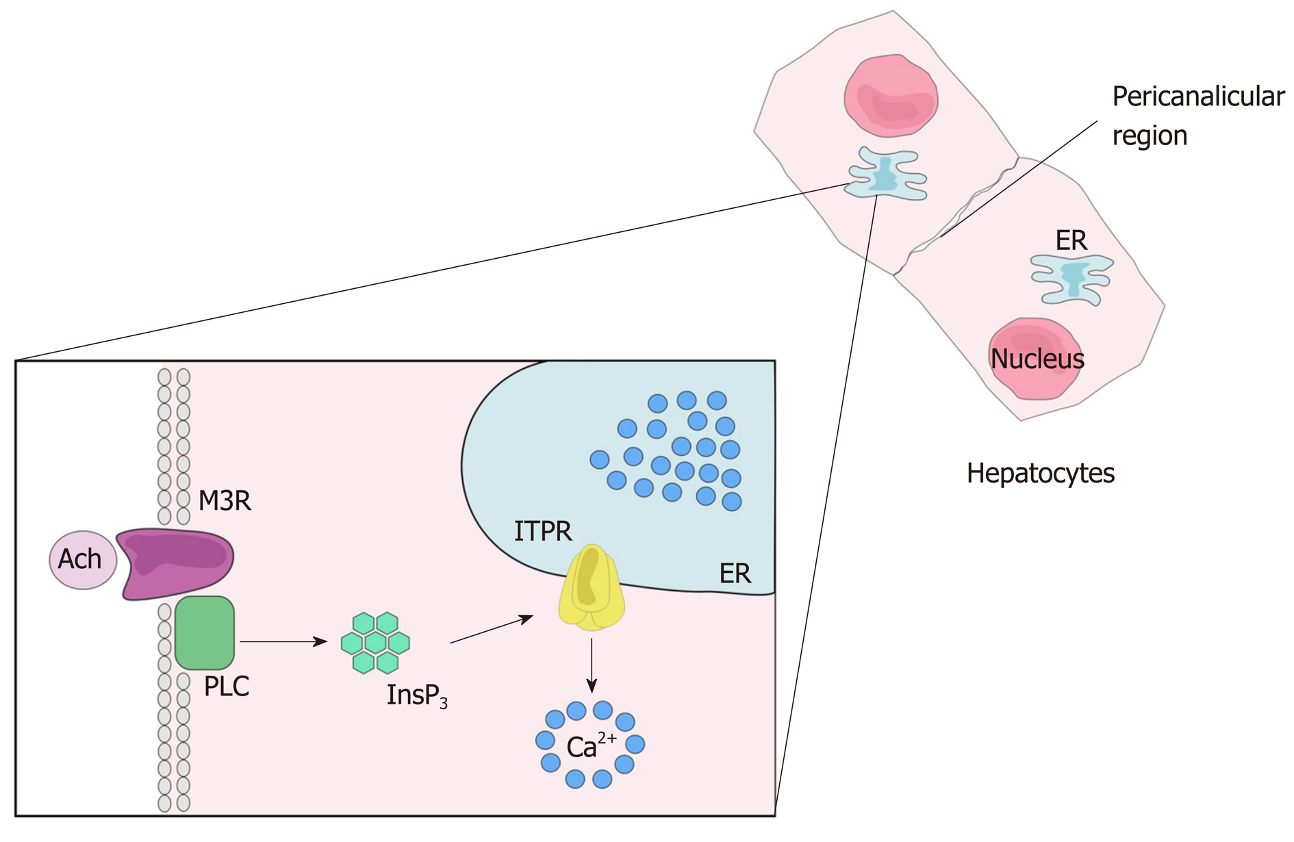
Figure 3 Calcium signaling.
After the ligation of an agonist to its receptor, here represent by the ligation of acetylcholine to muscarinic acetylcholine receptor, phospholipase C is activated and produces 1,4,5 inositol triphosphate. The inositol 1,4,5-trisphosphate binds its receptor, inositol 1,4,5-trisphosphate receptor, that is expressed mainly along the endoplasmic reticulum, leading to Ca2+ release into the cytosol. Ach: Acetylcholine; M3R: Muscarinic acetylcholine receptor; PLC: Phospholipase C; InsP3: Inositol 1,4,5-trisphosphate; ITPR: Inositol 1,4,5-trisphosphate receptor; ER: Endoplasmic reticulum.
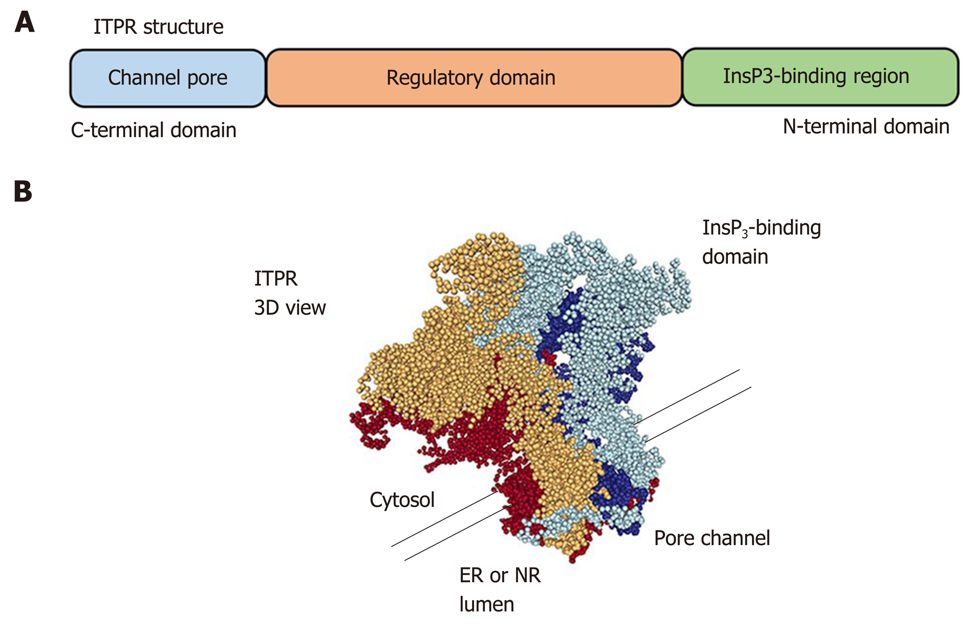
Figure 4 Inositol 1,4,5-trisphosphate receptor structure.
A: Linear structure of inositol 1,4,5-trisphosphate receptor (ITPR). The inositol 1,4,5-trisphosphate (InsP3)-binding domain is located on the N-terminal region and the pore channel is on the C-terminal region. The receptor spans organelle membrane six times; B: Tridimensional view of ITPR showing that the receptor is formed by a 4 single chain. It is necessary that four InsP3 molecules bind to the receptor to lead the calcium releases by a pore of the channel. The tridimensional structure was adapted from Molecular Modeling Database (National Center for Biotechnology Information). ER: Endoplasmic reticulum; ITPR: Inositol 1,4,5-trisphosphate receptor; NR: Nucleoplasmic reticulum.
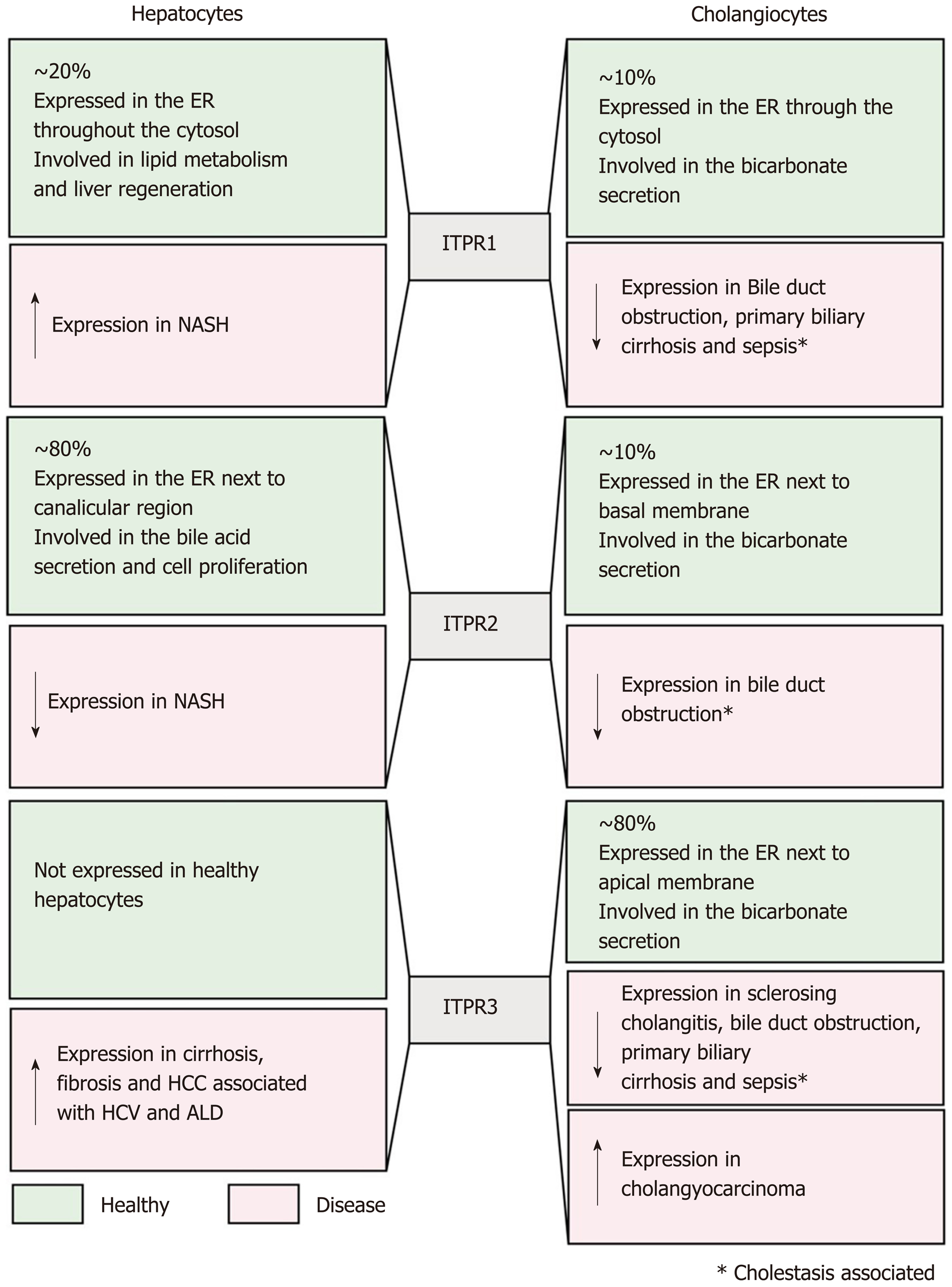
Figure 5 Inositol 1,4,5-trisphosphate receptors in the liver: Expression and functions.
This figure summarizes, in green, Inositol 1,4,5-trisphosphate receptor (ITPR) isoform expression in hepatocytes and cholangiocytes under physiological condition, and in red the expression level and function of each ITPR isoform in liver diseases. ITPR1: ITPR isoform 1; ITPR2: ITPR isoform 2; ITPR3: ITPR isoform 3; ER: Endoplasmic reticulum; NAFLD: Non-alcoholic fatty liver disease; HCC: Hepatocellular carcinoma.
- Citation: Lemos FO, Florentino RM, Lima Filho ACM, Santos MLD, Leite MF. Inositol 1,4,5-trisphosphate receptor in the liver: Expression and function. World J Gastroenterol 2019; 25(44): 6483-6494
- URL: https://www.wjgnet.com/1007-9327/full/v25/i44/6483.htm
- DOI: https://dx.doi.org/10.3748/wjg.v25.i44.6483


