Copyright
©The Author(s) 2019.
World J Gastroenterol. Nov 7, 2019; 25(41): 6248-6257
Published online Nov 7, 2019. doi: 10.3748/wjg.v25.i41.6248
Published online Nov 7, 2019. doi: 10.3748/wjg.v25.i41.6248
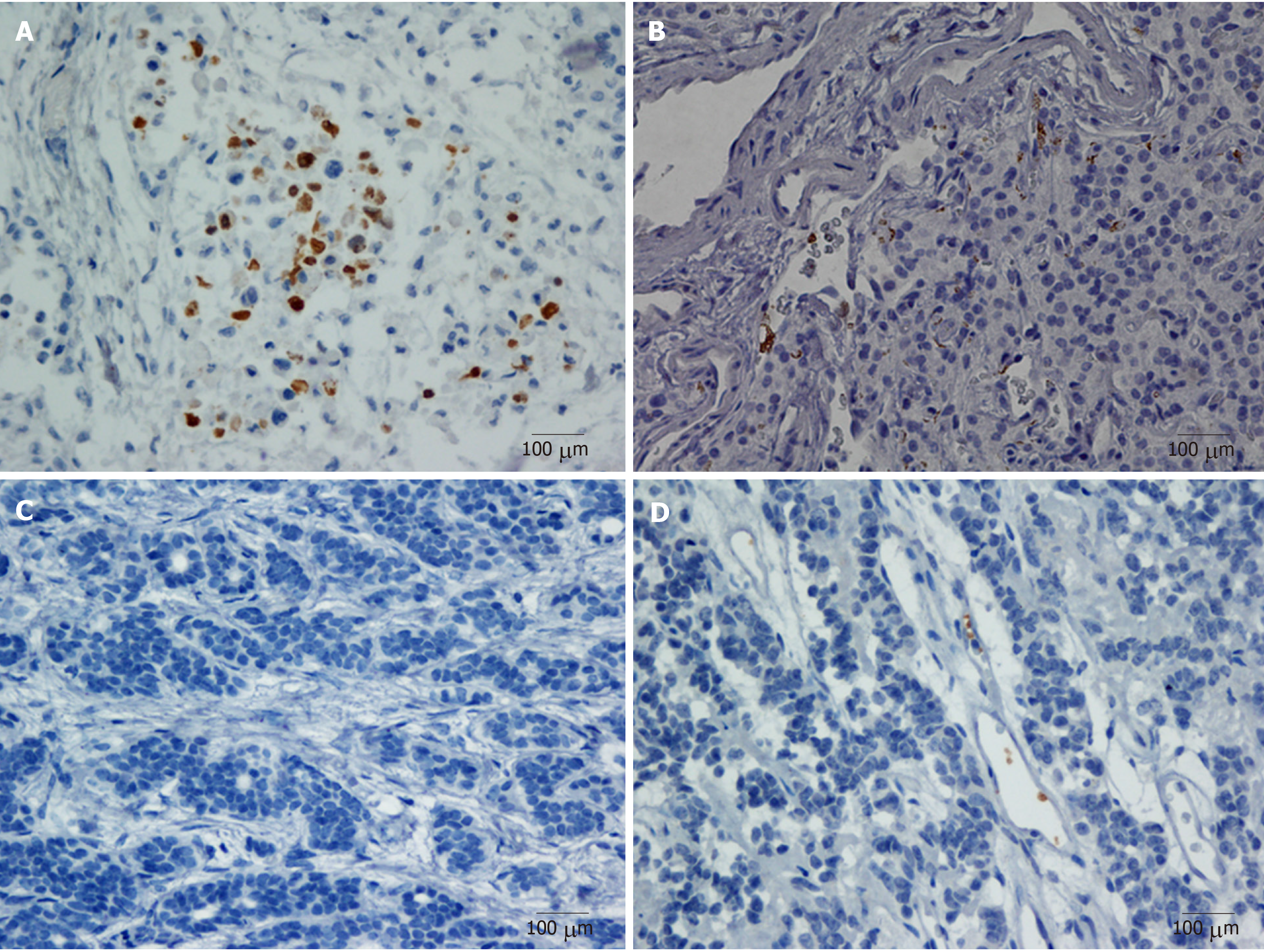
Figure 1 Representative microphotographs of CD42b staining.
A: Positive, CD42b expression aggregating around and embracing tumor cells; B: Positive, CD42b expression surrounding tumor cells and intratumoral blood vessels; C: Negative, no CD42b staining in intratumoral region; D: Negative, CD42b staining only in intratumoral vessels. All magnifications × 400. Positive staining appears brown.
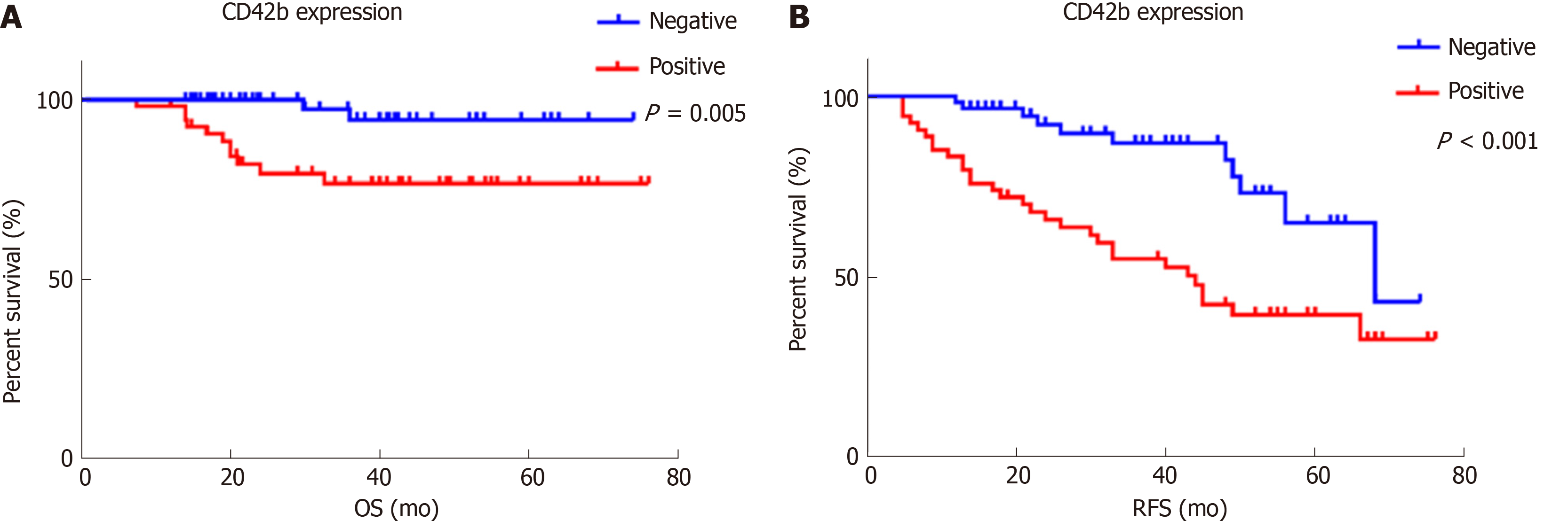
Figure 2 The relationship between CD42b expression and overall survival and recurrence-free survival.
A: OS (aP = 0.005); B: RFS (bP < 0.001). Patients with positive CD42b expression had worse OS and RFS than those with negative CD42b expression. P values were calculated by Kaplan–Meier estimations. OS: Overall survival; RFS: Recurrence-free survival.
- Citation: Xu SS, Xu HX, Wang WQ, Li S, Li H, Li TJ, Zhang WH, Liu L, Yu XJ. Tumor-infiltrating platelets predict postoperative recurrence and survival in resectable pancreatic neuroendocrine tumor. World J Gastroenterol 2019; 25(41): 6248-6257
- URL: https://www.wjgnet.com/1007-9327/full/v25/i41/6248.htm
- DOI: https://dx.doi.org/10.3748/wjg.v25.i41.6248


