Copyright
©The Author(s) 2019.
World J Gastroenterol. Oct 14, 2019; 25(38): 5883-5896
Published online Oct 14, 2019. doi: 10.3748/wjg.v25.i38.5883
Published online Oct 14, 2019. doi: 10.3748/wjg.v25.i38.5883
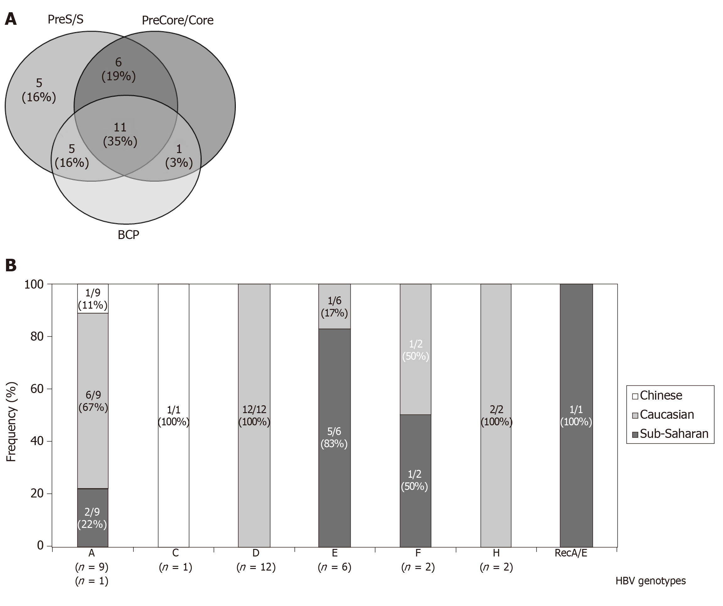
Figure 1 Features of samples analyzed.
A: Number of patients with HBV-DNA amplification positive results in the different genomic regions analysed; B: Ethnic group distribution among the different HBV genotypes. The 4 patients in whom were confirmed sequential changes of the genotyping results during the follow-up were included in both genotype categories. BCP: Basal core promoter; HBV: Hepatitis B virus.
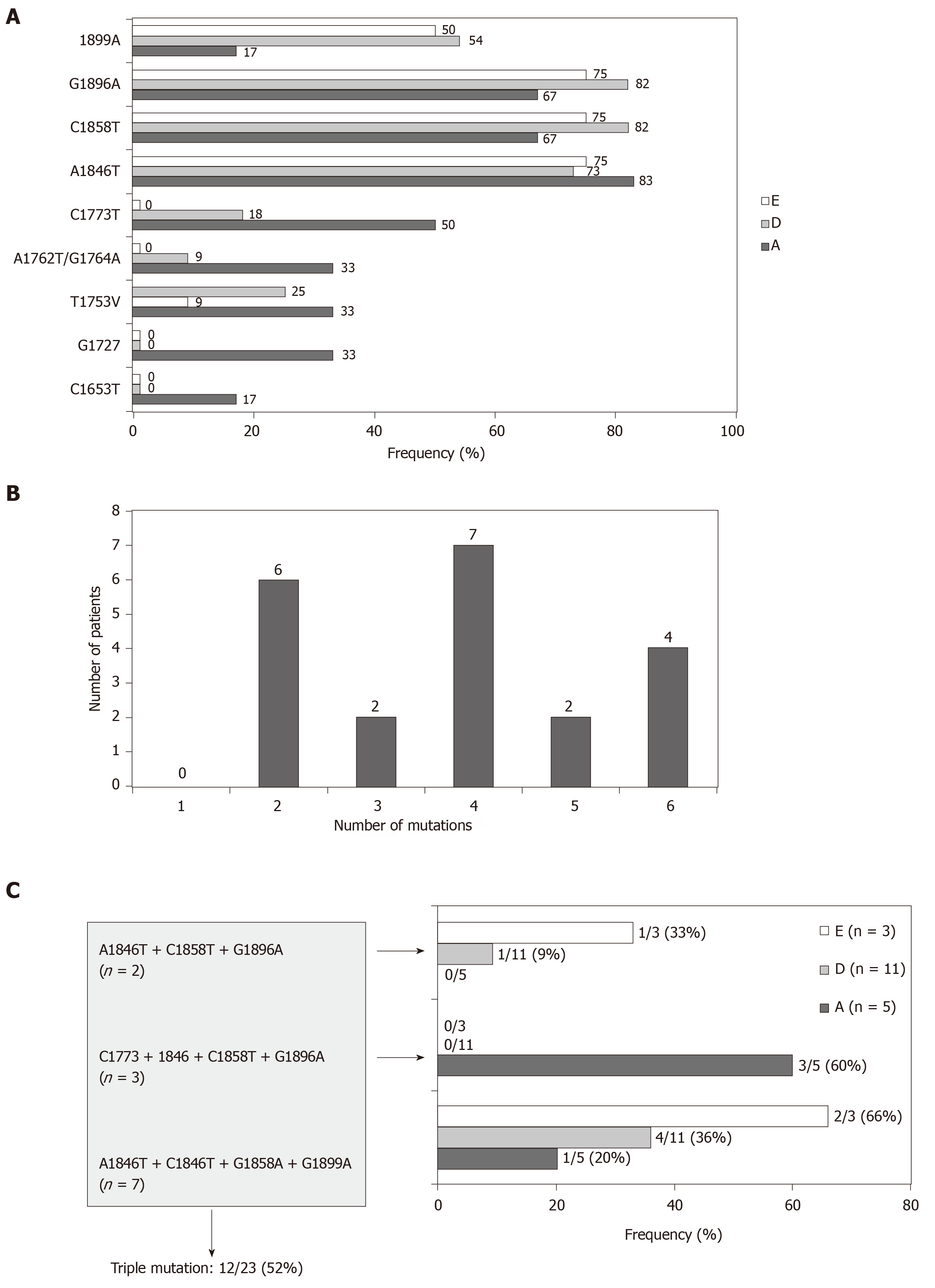
Figure 2 Analysis of basal core promoter /precore/core mutants related with the e-antigen negative status.
A: Prevalence of individual mutations of the in the 3 most frequent HBV genotypes analyzed; B: Frequency of coexistence of more than one BCP/precore/core mutants; and C: Frequency of the most common combination of mutations in the different genotypes. The frequency calculation was performed including only the 23 patients with precore/core sequences data, not in on intention to treat basis. BCP: Basal core promoter; HBV: Hepatitis B virus; A: HBV-A; D: HBV-D; E: HBV-E.
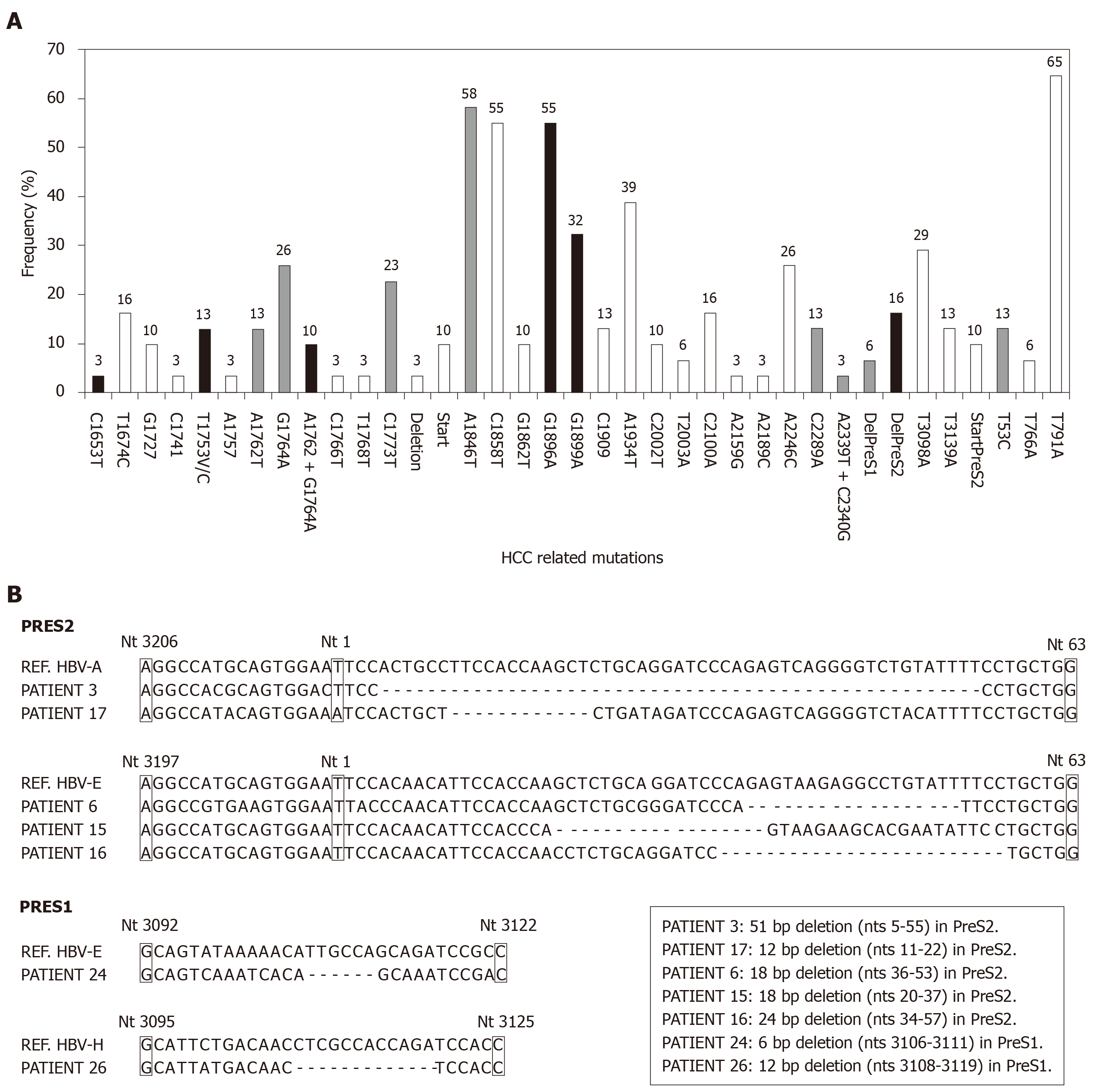
Figure 3 Analysis of basal core promoter/precore/core region mutants related with the hepatocellular carcinoma risk.
A: Frequency distribution of BCP/precore/core and HBsAg mutations (Black, grey and white bars show the viral variants with the highest, median and lowest evidences of association with HCC, respectively); B: Sequences of the 7 patients with preS deletions. BCP: Basal core promoter; HCC: Hepatocellular carcinoma.
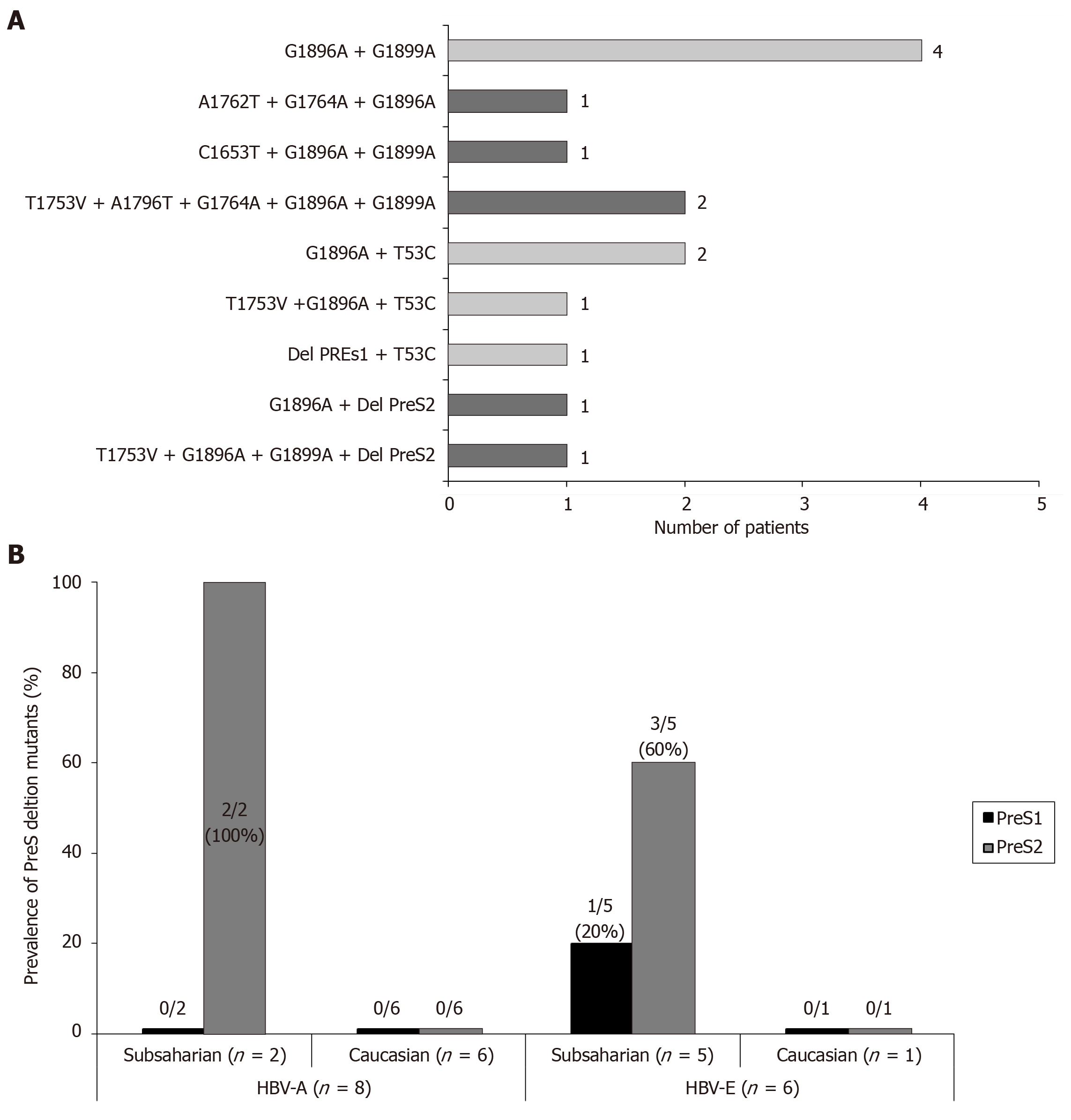
Figure 4 Analysis of preS deletion mutations related with the hepatocellular carcinoma risk.
A: Frequency of coexistence of hepatocellular carcinoma related mutations in both BCP/precore and preS regions (Dark bars indicate the patients with theoretical highest risk of hepatocellular carcinoma development, based in the coexistence of mutations in the HBsAg and precore region, or the simultaneous detection of 3 or more mutation irrespective of the genomic region); B: Prevalence of preS deletions in different genotypes and ethnic groups. Only a Caucasian patient infected with HBV-H genotype had a preS1 deletion. BCP: Basal core promoter.
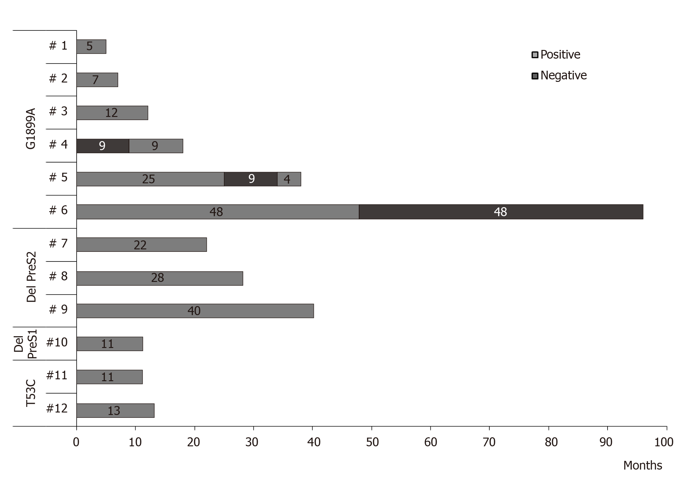
Figure 5 Persistence over time of the high-risk mutations as major population in the patients analyzed.
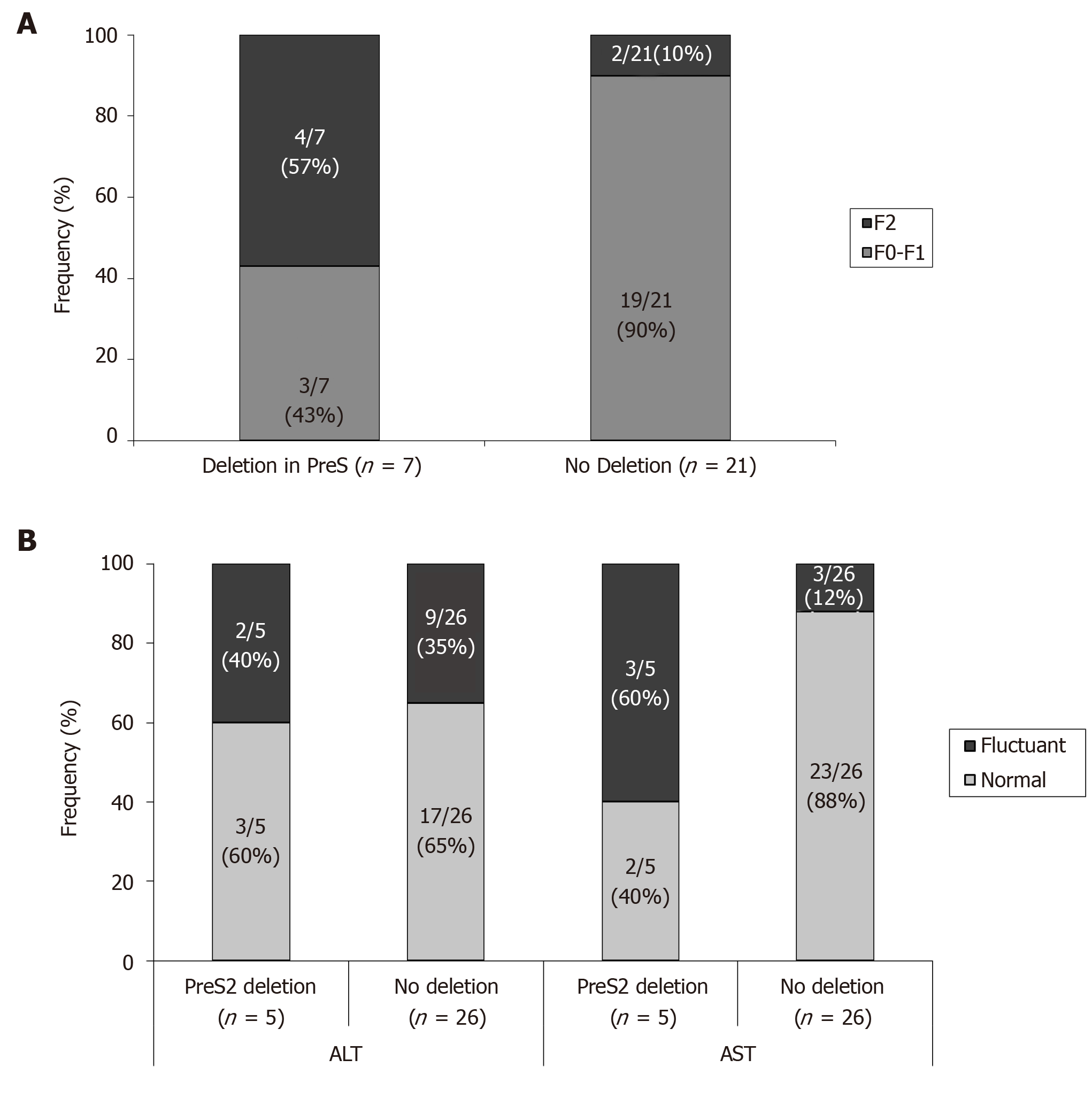
Figure 6 Differences between patients with or without preS deletion mutants.
A: Differences between patients with or without preS deletion mutants at fibrosis level; B: Differences between patients with or without preS deletion mutants at evolution of transaminase levels during follow-up. AST: Aspartate aminotransferase; ALT: Alanine aminotransferase.
- Citation: Gil-García AI, Madejón A, Francisco-Recuero I, López-López A, Villafranca E, Romero M, García A, Olveira A, Mena R, Larrubia JR, García-Samaniego J. Prevalence of hepatocarcinoma-related hepatitis B virus mutants in patients in grey zone of treatment. World J Gastroenterol 2019; 25(38): 5883-5896
- URL: https://www.wjgnet.com/1007-9327/full/v25/i38/5883.htm
- DOI: https://dx.doi.org/10.3748/wjg.v25.i38.5883


