Copyright
©The Author(s) 2019.
World J Gastroenterol. Aug 28, 2019; 25(32): 4580-4597
Published online Aug 28, 2019. doi: 10.3748/wjg.v25.i32.4580
Published online Aug 28, 2019. doi: 10.3748/wjg.v25.i32.4580
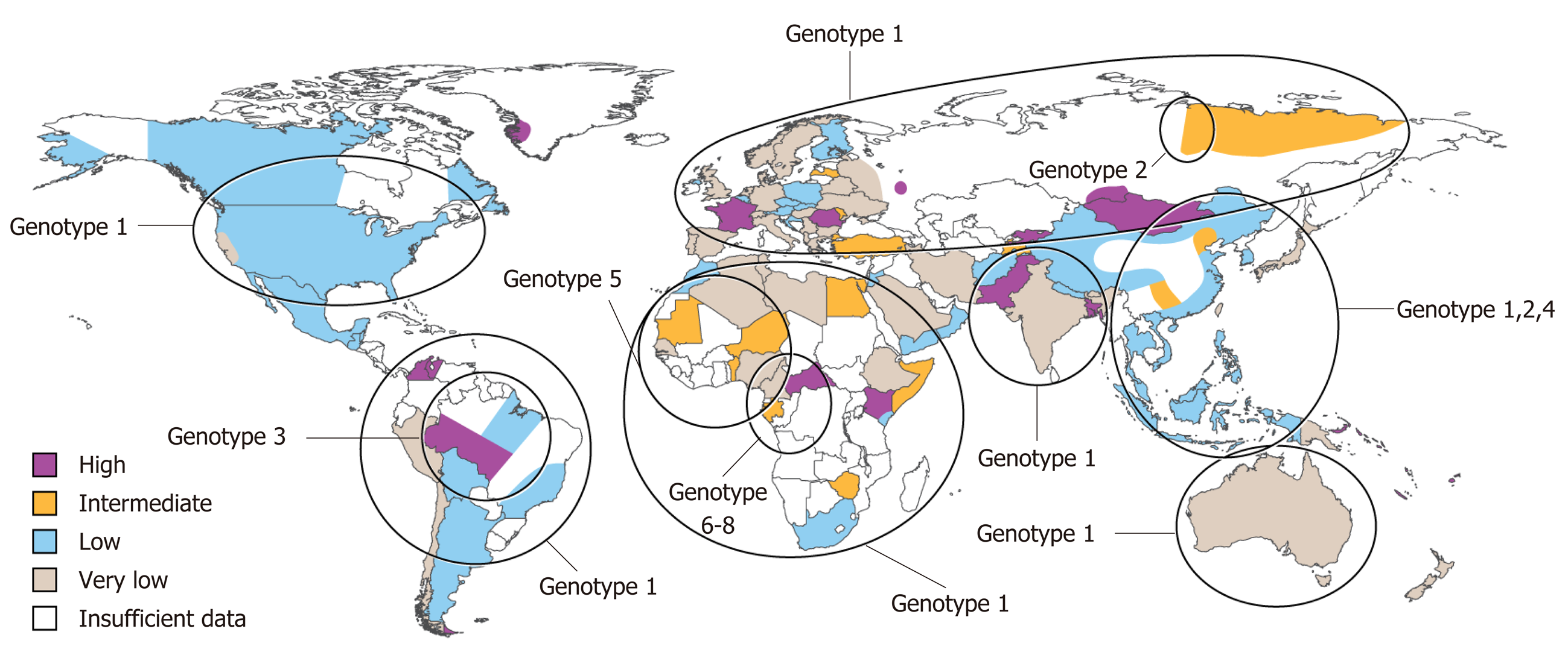
Figure 1 World map illustrating the estimated prevalence of hepatitis D virus infection with genotypic distribution.
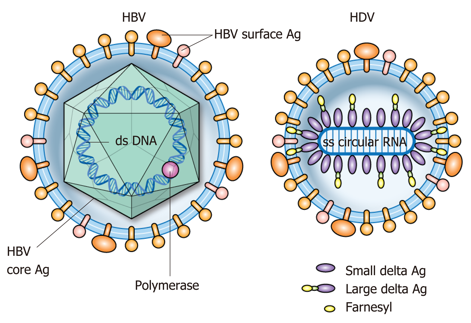
Figure 2 Structural representation of hepatitis B and delta viruses.
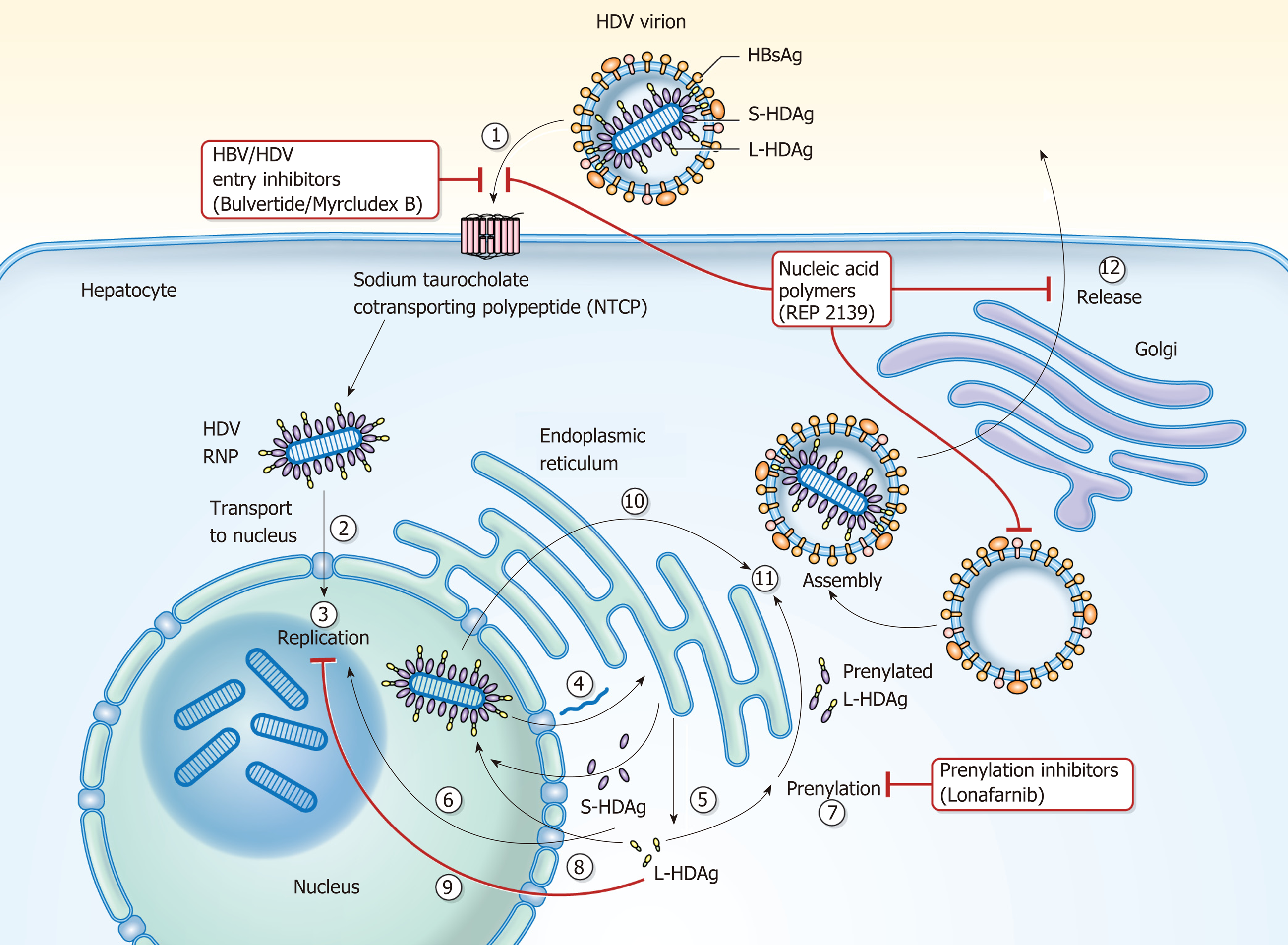
Figure 3 Hepatitis D virus viral life cycle and sites of investigative therapies.
(1) Hepatitis D virus (HDV) virion attaches to the hepatocyte through interaction between HBsAg and NTCP; (2) HDV RNP is translocated to nucleus facilitated by HDAg; (3) HDV genome replication occurs via a “rolling cycle” mechanism; (4) HDV antigenome is transported out of the nucleus to the endoplasmic reticulum (ER); (5) HDV antigenome is translated in the ER into SHDAg and LHDAg; (6) SHDAg is transported into the nucleus; (7) SHDAg promotes HDV replication in the nucleus; (8) LHDAg undergoes prenylation prior to assembly; (9) LHDAg inhibits HDV replication in the nucleus; (10) New HDAg molecules are associated with new transcripts of genomic RNA to form new RNPs that are exported to the cytoplasm; (11) New HDV RNPs associate with HBsAg and assemble into HDV virions; and (12) Completed HDV virions are released from the hepatocyte via the trans-Golgi network.
- Citation: Gilman C, Heller T, Koh C. Chronic hepatitis delta: A state-of-the-art review and new therapies. World J Gastroenterol 2019; 25(32): 4580-4597
- URL: https://www.wjgnet.com/1007-9327/full/v25/i32/4580.htm
- DOI: https://dx.doi.org/10.3748/wjg.v25.i32.4580


