Copyright
©The Author(s) 2019.
World J Gastroenterol. Aug 7, 2019; 25(29): 3857-3869
Published online Aug 7, 2019. doi: 10.3748/wjg.v25.i29.3857
Published online Aug 7, 2019. doi: 10.3748/wjg.v25.i29.3857
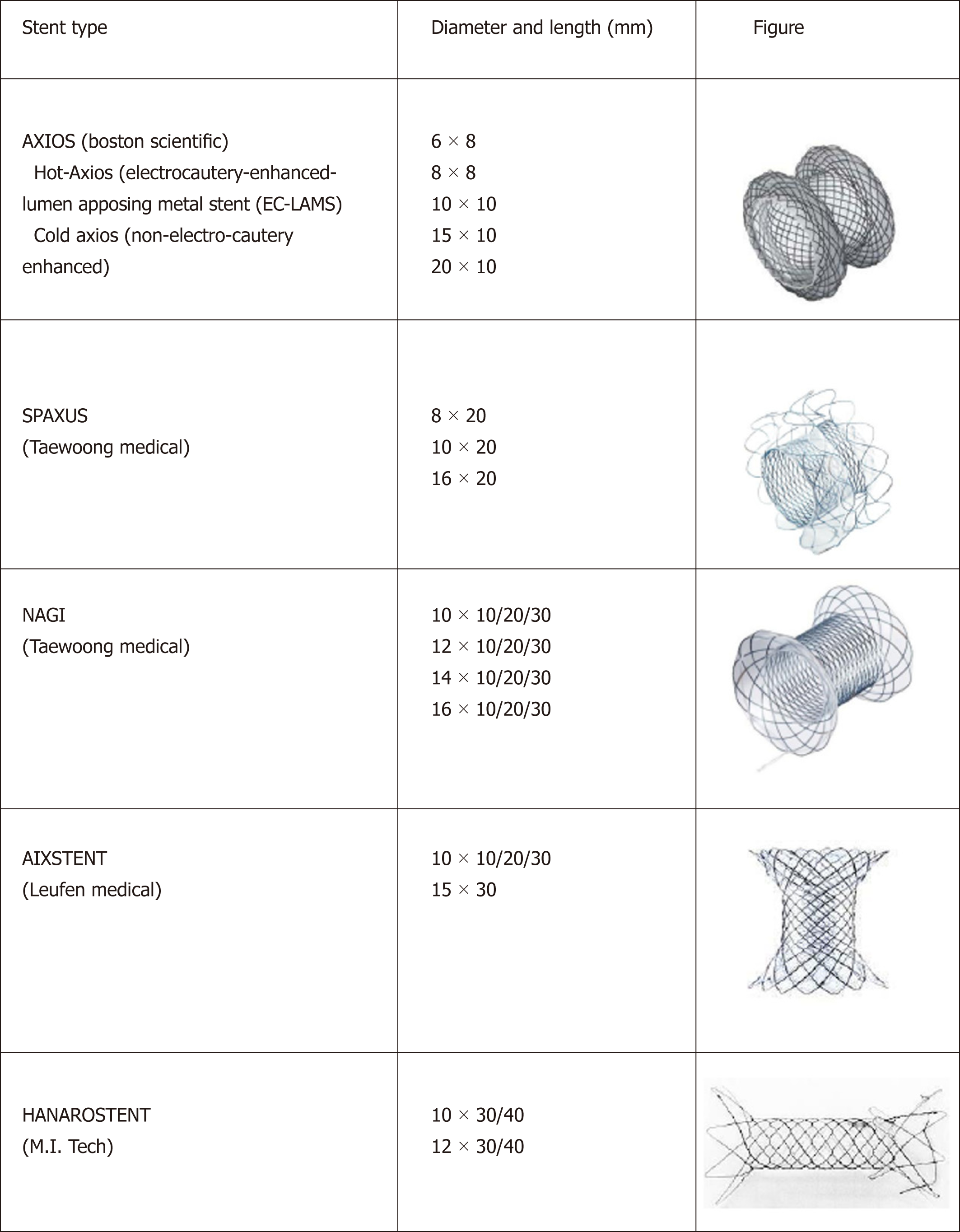
Figure 1 Currently lumen-apposing metal stent and fully covered self-expanding metal stent with peculiar anti-migratory shape available on the market.
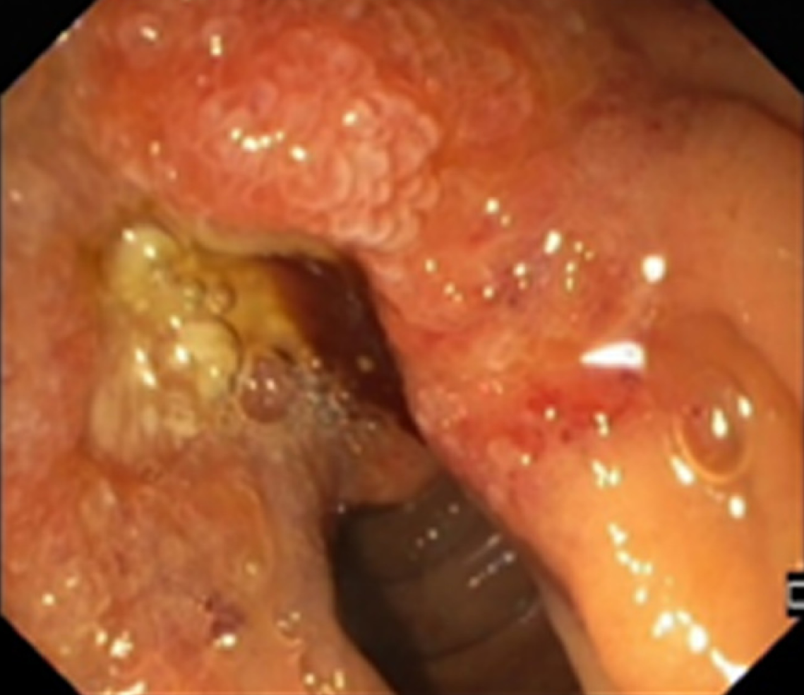
Figure 2 Endoscopic view of infiltration of the papilla by invasive pancreatic cancer.
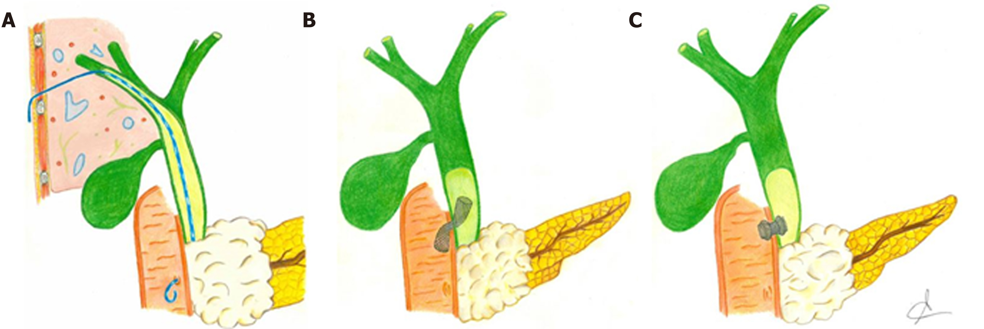
Figure 3 Graphic representation of the main interventional techniques applied to perform biliary drainage after failed endoscopic retrograde cholangiopancreatography in malignant biliary obstruction.
A: Percutaneous transhepatic biliary drainage; B: Endoscopic ultrasonography-guided choledocho-duodenostomy with placement of bilary fully covered self-expanding metal stent; C: Endoscopic ultrasonography-guided choledocho-duodenostomy with placement of lumen-apposing metal stent.
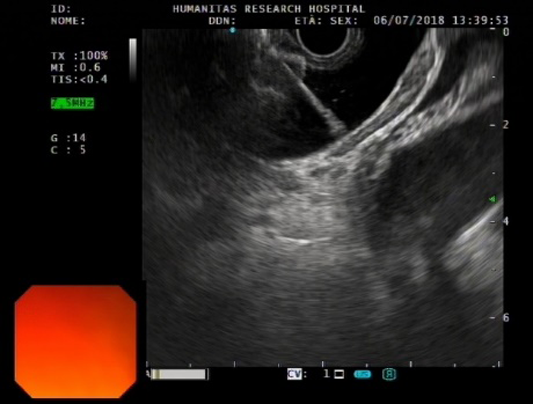
Figure 4 Echoendoscopic view of the first flange deployment of electrocautery-enhanced lumen-apposing metal stent in a dilated common bile duct.
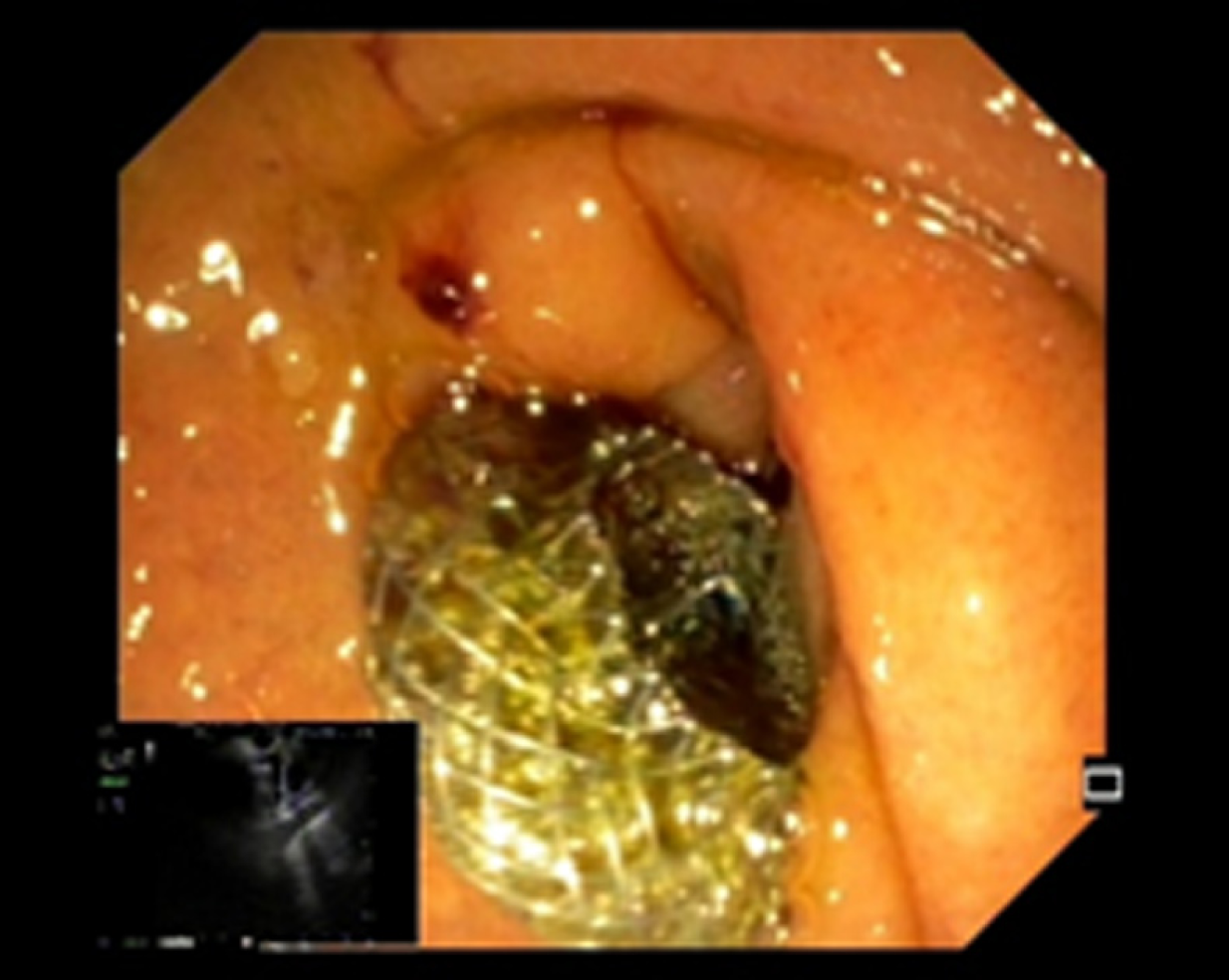
Figure 5 Final endoscopic appearance of electrocautery-enhanced lumen-apposing metal stent deployed in the duodenal bulb.
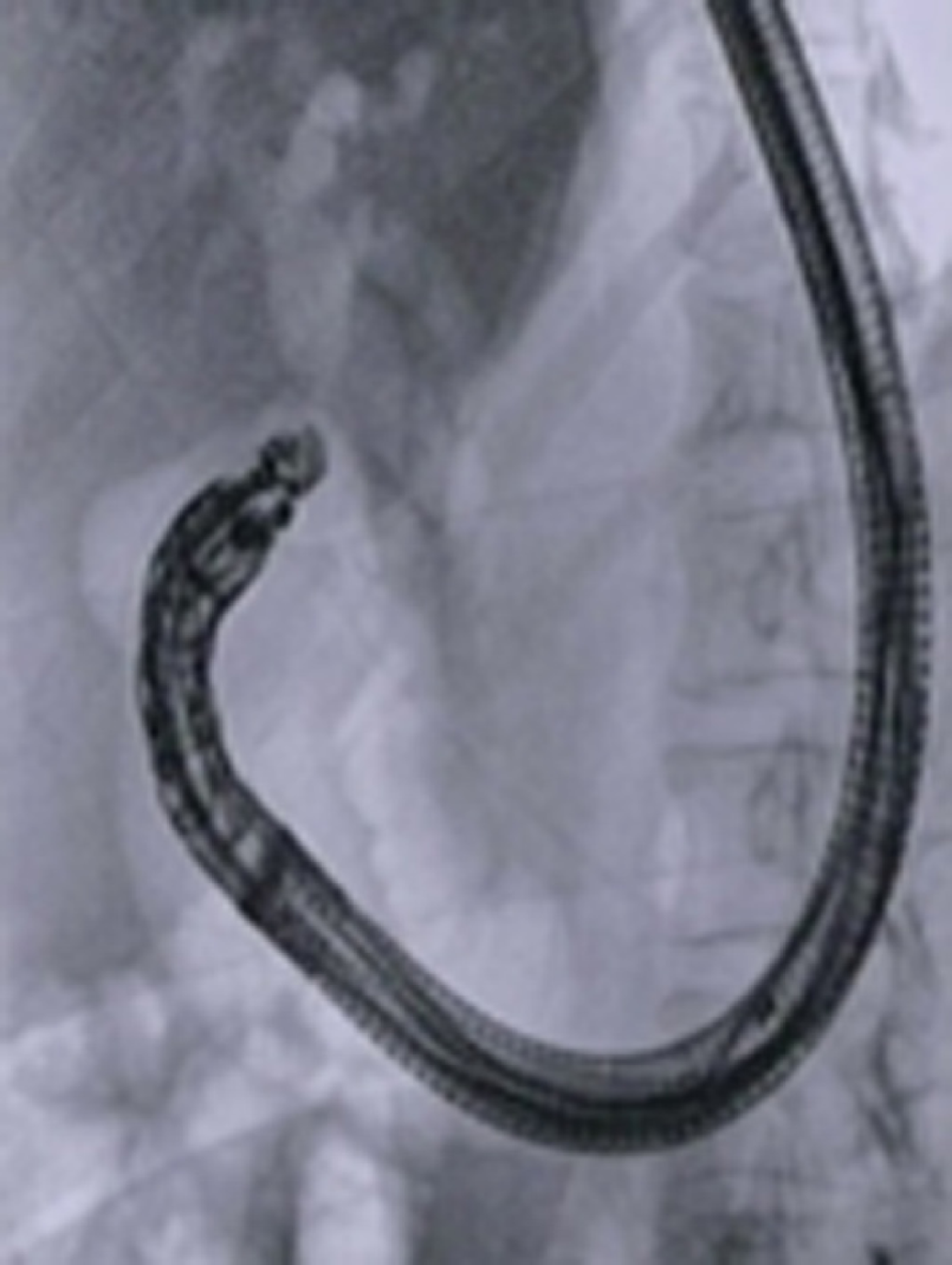
Figure 6 Final RX appearance of electrocautery-enhanced lumen-apposing metal stent deployed across the duodenal bulb into the common bile duct.
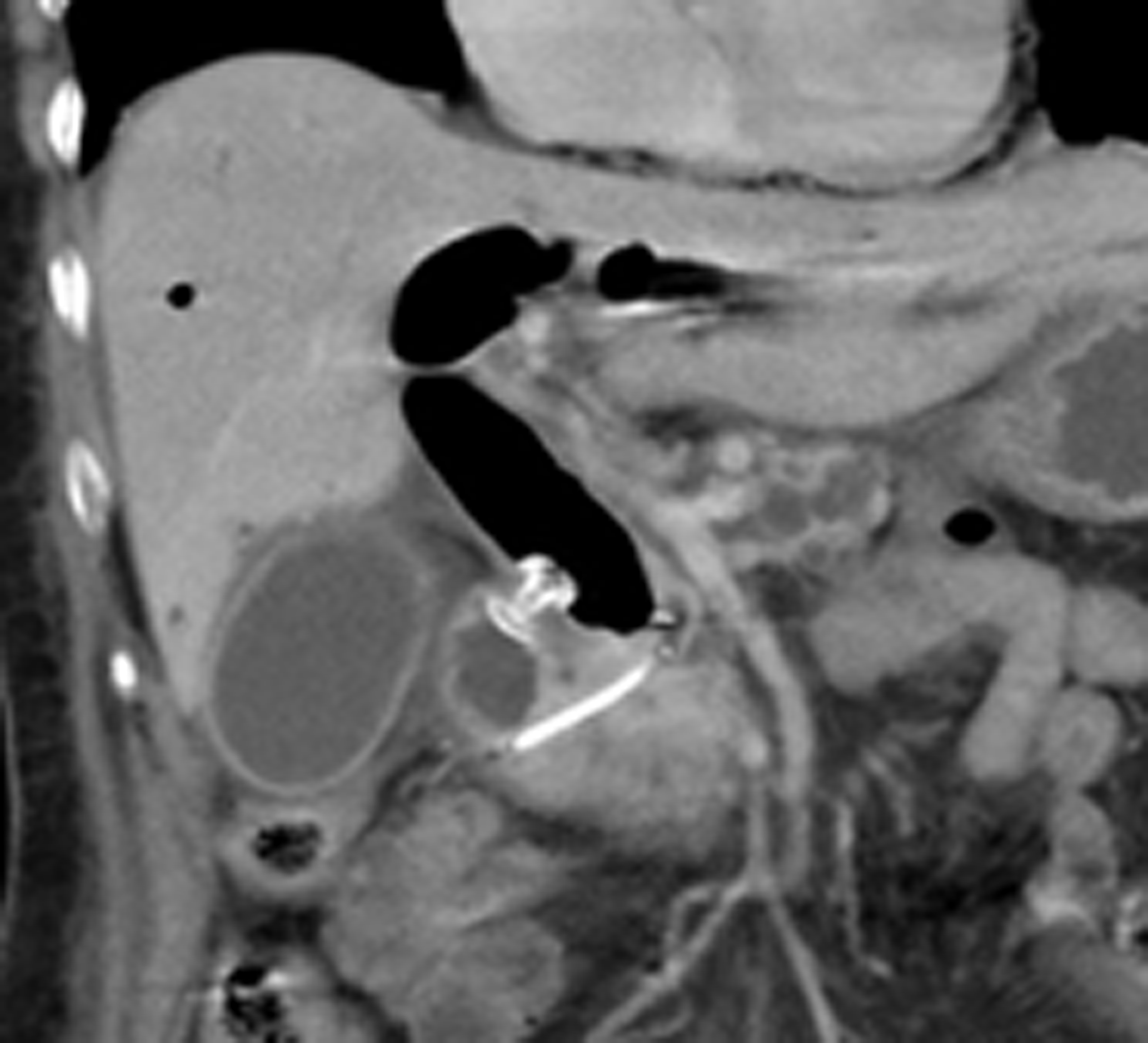
Figure 7 Computed tomography scan appearance of electrocautery-enhanced lumen-apposing metal stent deployed across the duodenal bulb and plastic pancreatic stent previously placed during failed endoscopic retrograde cholangiopancreatography.
- Citation: Anderloni A, Troncone E, Fugazza A, Cappello A, Del Vecchio Blanco G, Monteleone G, Repici A. Lumen-apposing metal stents for malignant biliary obstruction: Is this the ultimate horizon of our experience? World J Gastroenterol 2019; 25(29): 3857-3869
- URL: https://www.wjgnet.com/1007-9327/full/v25/i29/3857.htm
- DOI: https://dx.doi.org/10.3748/wjg.v25.i29.3857


