Copyright
©The Author(s) 2018.
World J Gastroenterol. Dec 21, 2018; 24(47): 5331-5337
Published online Dec 21, 2018. doi: 10.3748/wjg.v24.i47.5331
Published online Dec 21, 2018. doi: 10.3748/wjg.v24.i47.5331
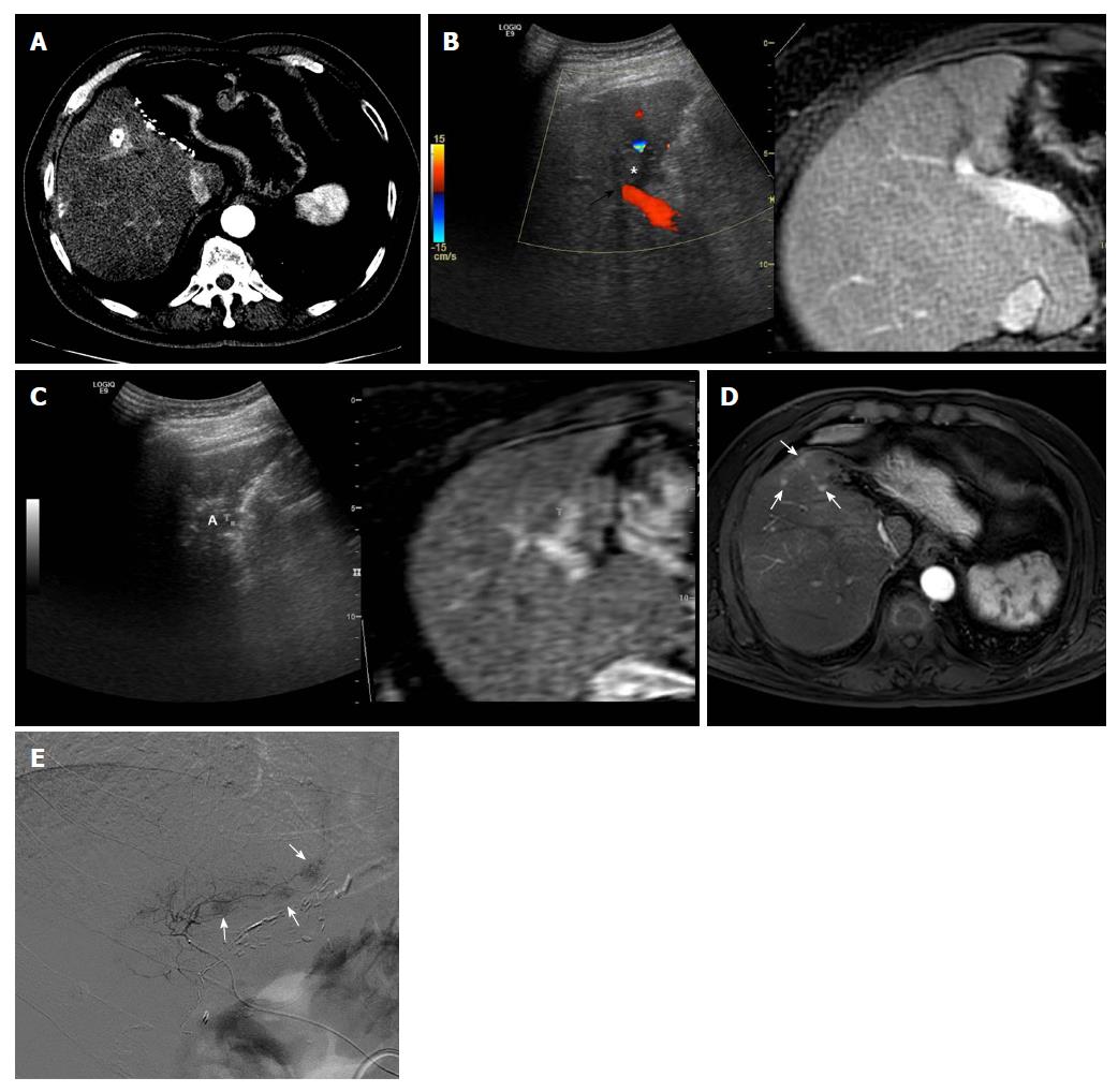
Figure 1 Images demonstrating aggressive intrasegmental recurrence after radiofrequency ablation for perivascular hepatocellular carcinoma.
A: Axial computed tomography image obtained during hepatic arterial phase shows viable hepatocellular carcinoma (HCC) within the partially lipiodolized nodule (asterisk) in segment V before radiofrequency (RF) ablation. The index tumor is in contact with the right portal vein (black arrow); B: On planning ultrasonography (US), using fusion imaging with color Doppler US and magnetic resonance imaging (MRI), the low echogenic incident tumor (asterisk) is in contact with a right portal vein (black arrow); C: During RF ablation with the US fusion system, the ablation zone (A) is covered with viable enhancing tumor foci, indicating T marker on real time US/fused MR image; D: MRI scan obtained during the hepatic arterial phase 9 mo after RF ablation shows multiple small arterial enhancing nodules (white arrows) of consistent size, representing recurrent tumors. These recurrent tumors developed simultaneously in a peripheral area of the treated segment, fed by the previous peritumoral portal vein; E: The patient underwent transarterial chemoembolization for tumor control considering tumor multiplicity. Multiple small nodular tumors were detected along the portal tract on hepatic angiogram.
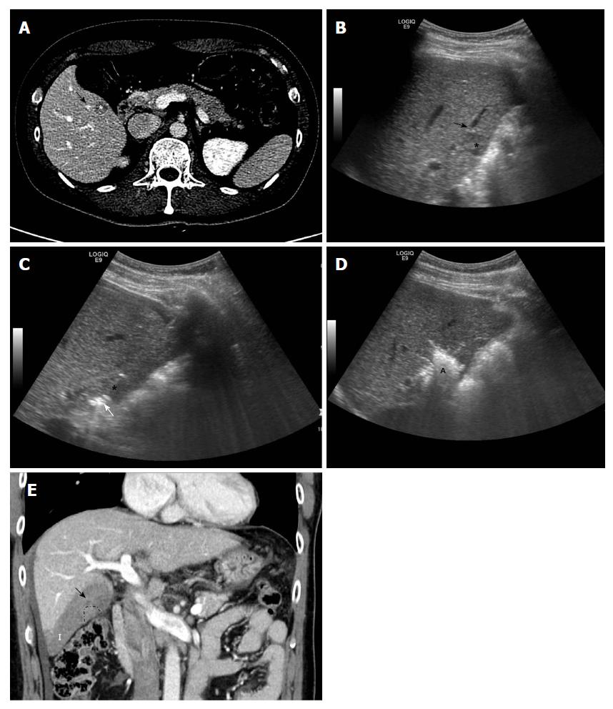
Figure 2 Images showing subsegmental hepatic infarction after radiofrequency ablation for perivascular hepatocellular carcinoma.
A: Axial computed tomography image obtained during equilibrium phase shows 1.3-cm hepatocellular carcinoma (asterisk) in segment V before radiofrequency (RF) ablation. The index tumor is in contact with the right portal vein (black arrow); B: Planning ultrasound image obtained before RF ablation shows the low-echoic-index tumor (asterisk) in contact with a right portal vein (black arrow); C: During RF ablation, the RF electrode (white arrow) is inserted into the index tumor (asterisk), evading the adjacent portal vein; D: At the end of the procedure, a hyperechoic ablation zone (A) completely covered the index tumor; E: Thrombosis within the peritumoral portal vein (black arrow) developed around the index tumor (dotted line), shown on coronal computed tomography images obtained immediately after RF ablation. This led to subsegmental infarction (I) in the peripheral area of hepatic segment VI.
- Citation: Kang TW, Lim HK, Cha DI. Percutaneous ablation for perivascular hepatocellular carcinoma: Refining the current status based on emerging evidence and future perspectives. World J Gastroenterol 2018; 24(47): 5331-5337
- URL: https://www.wjgnet.com/1007-9327/full/v24/i47/5331.htm
- DOI: https://dx.doi.org/10.3748/wjg.v24.i47.5331


