Copyright
©The Author(s) 2018.
World J Gastroenterol. Oct 7, 2018; 24(37): 4272-4280
Published online Oct 7, 2018. doi: 10.3748/wjg.v24.i37.4272
Published online Oct 7, 2018. doi: 10.3748/wjg.v24.i37.4272
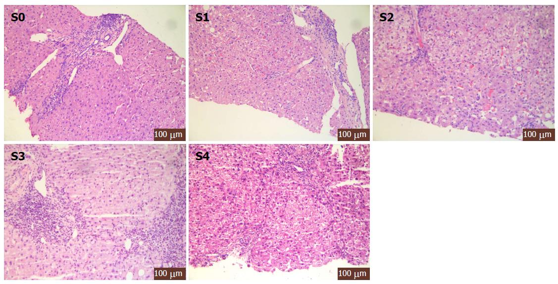
Figure 1 Histopathology of different pathological stages of hepatic fibrosis.
S0 phase: No fibrosis; S1 phase: Fibrous enlargement in the portal area, but no fibrillary septum formation; S2 phase: Fibrosis enlargement in the portal area, a few fibrous septae formed; S3 stage: Most fibrous septae formed but no hardened nodules; S4 stage: Liver cirrhosis.
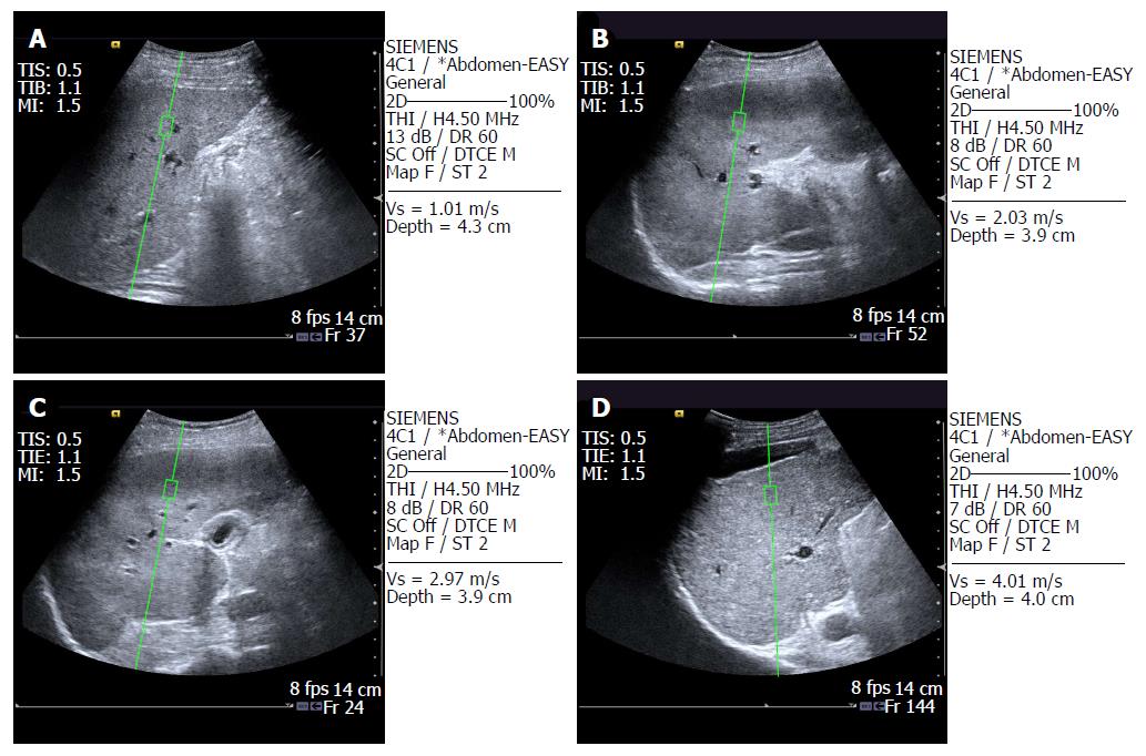
Figure 2 Image of hepatic fibrosis assessed by acoustic radiation force impulse to assess liver tissue elasticity.
A: Normal liver tissue, SWV = 1.01 m/s; B: Mild hepatic fibrosis, SWV = 2.03 m/s; C: Moderate hepatic fibrosis, SWV = 2.97 m/s; D: Severe hepatic fibrosis, SWV = 4.01m/s. ARFI: Acoustic radiation force impulse.
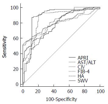
Figure 3 Receiver operating characteristic curve for the diagnosis of hepatic fibrosis based on different indicators.
APRI: Aspartate aminotransferase-to-platelet ratio index; AST: Aspartate transaminase; ALT: Alanine aminotransferase; CIV: Type IV collagen; FIB-4: Fibrosis index based on the 4 factor; HA: Hyaluronic acid; SWV: Shear wave velocity.
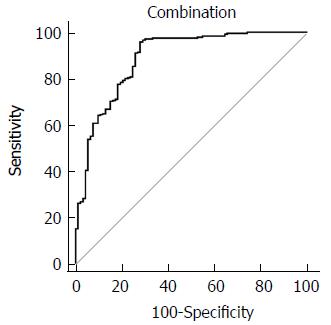
Figure 4 Receiver operating characteristic curve for the diagnosis of hepatic fibrosis based on different combined indicators.
- Citation: Xu B, Zhou NM, Cao WT, Li XJ. Evaluation of elastography combined with serological indexes for hepatic fibrosis in patients with chronic hepatitis B. World J Gastroenterol 2018; 24(37): 4272-4280
- URL: https://www.wjgnet.com/1007-9327/full/v24/i37/4272.htm
- DOI: https://dx.doi.org/10.3748/wjg.v24.i37.4272


