Copyright
©The Author(s) 2018.
World J Gastroenterol. Sep 7, 2018; 24(33): 3760-3769
Published online Sep 7, 2018. doi: 10.3748/wjg.v24.i33.3760
Published online Sep 7, 2018. doi: 10.3748/wjg.v24.i33.3760
Figure 1 Metabolite-concentration distribution of gastric cancer tumor and its surrounding healthy tissue using the partial least squares discriminate analysis statistical method.
A and B: Tumor (green, n = 19) vs healthy tissue (red, n = 19); C: and D: Tumor between chromosomal instability (CIN) type (red, n = 9) and non-CIN type (green, n = 10); E and F: Tumor (green, n = 9) and healthy tissue (red, n = 9) in the CIN type; G and H: Tumor (green, n = 10) and healthy tissue (red, n = 10) in the non-CIN type. A/C/E/G and B/D/F/H defined metabolites from ESI- and ESI+ LC-MS. Arrows indicate the variation in the two groups of metabolite distribution. The straight line indicates the difference of metabolite distribution between two patient types. Within the ellipse was the 95% confidence region. CIN: Chromosomal instability; ESI: Electrospray ionization; LC-MS: Liquid chromatography-mass spectrometry.
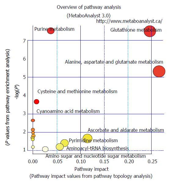
Figure 2 Pathways in chromosomal instability (CIN) and non-chromosomal instability gastric cancer.
Pathways involving glutathione metabolism, aminoacyl-tRNA biosynthesis, arginine and proline metabolism, and glutamine and glutamate metabolism were found in CIN and non-CIN types. Pathways involving alanine, aspartate, and glutamate metabolism, glyoxylate and dicarboxylate metabolism, histidine metabolism, and phenylalanine, tyrosine, and tryptophan biosynthesis were observed in the CIN type but not the non-CIN type. CIN: Chromosomal instability.
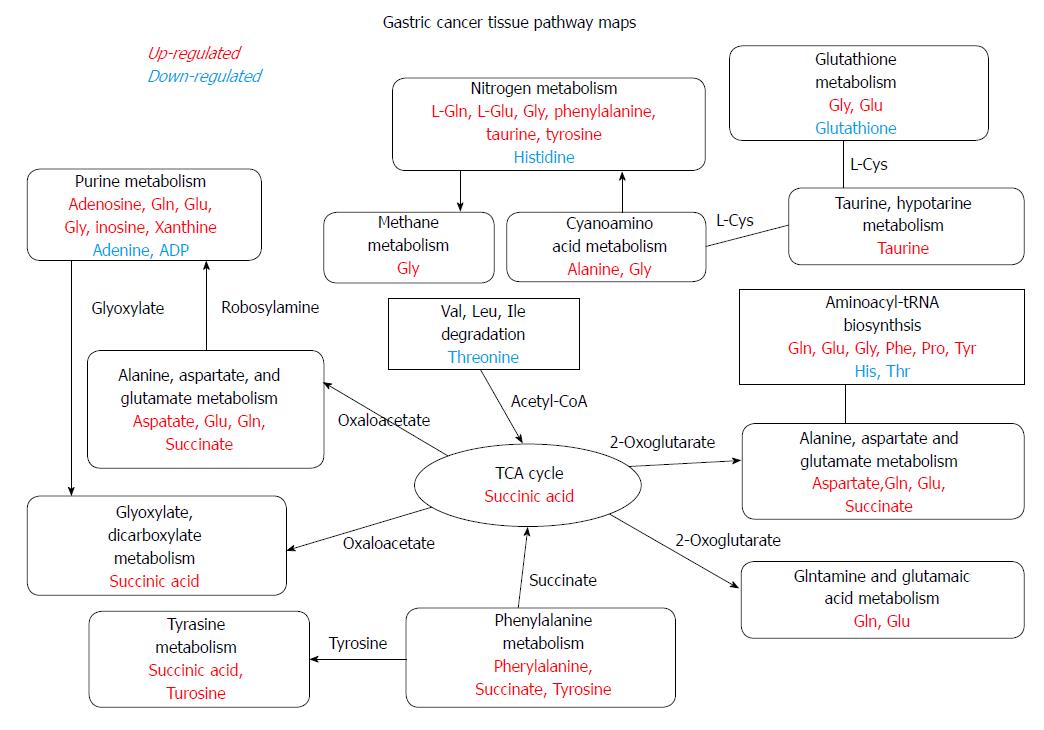
Figure 3 Metabolites in the chromosomal instability type and the non-chromosomal instability type.
Upregulated (red) or down-regulated (blue) metabolites of pathways (black) involved in CIN and non-CIN types. CIN: Chromosomal instability.
- Citation: Tsai CK, Yeh TS, Wu RC, Lai YC, Chiang MH, Lu KY, Hung CY, Ho HY, Cheng ML, Lin G. Metabolomic alterations and chromosomal instability status in gastric cancer. World J Gastroenterol 2018; 24(33): 3760-3769
- URL: https://www.wjgnet.com/1007-9327/full/v24/i33/3760.htm
- DOI: https://dx.doi.org/10.3748/wjg.v24.i33.3760


