Copyright
©The Author(s) 2018.
World J Gastroenterol. May 7, 2018; 24(17): 1888-1900
Published online May 7, 2018. doi: 10.3748/wjg.v24.i17.1888
Published online May 7, 2018. doi: 10.3748/wjg.v24.i17.1888
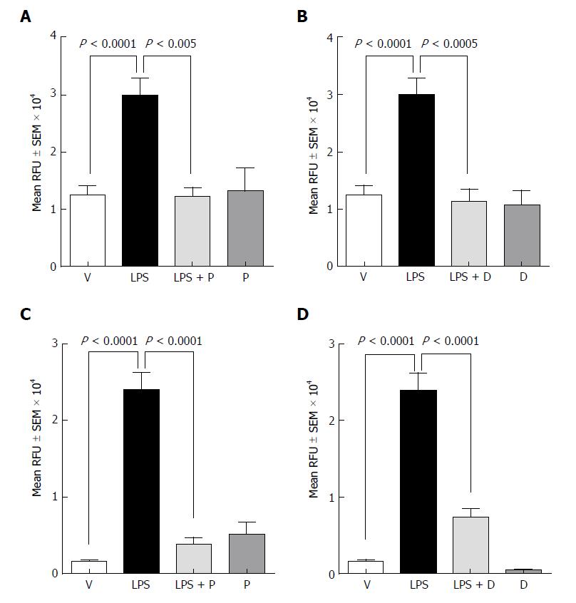
Figure 1 Effect of plecanatide and dolcanatide on lipopolysaccharide-induced increase in permeability of 4 kDa fluorescein isothiocyanate-dextran across Caco-2 and T84 cell monolayers.
Caco-2 (A and B) and T84 (C and D) cells cultured on snap well inserts were treated with vehicle or 1 μmol/L of plecanatide (A and C) or dolcanatide (B and D) in the presence or absence of 100 μg/mL of LPS for 16 h. Subsequently, 1 mg/mL of FITC-dextran was added to the apical compartment. Paracellular permeability was determined by measuring the amount of FITC-dextran present in the basal compartment. Data represent mean ± SEM analyzed in triplicates. D: Dolcanatide; FITC: Fluorescein isothiocyanate; LPS: Lipopolysaccharide; P: Plecanatide; RFU: Relative fluorescence units; SEM: Standard error of the mean; V: Vehicle.
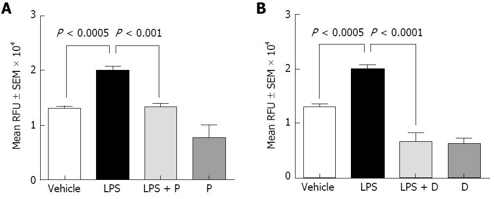
Figure 2 Effect of plecanatide (A) and dolcanatide (B) on lipopolysaccharide-induced increased permeability of 4 kD fluorescein isothiocyanate-dextran across rat colon tissues.
Rat colon tissues (2 cm pieces) were incubated overnight with vehicle, 10 μM plecanatide or dolcanatide in the presence or absence of 100 μg/mL LPS. Data represent mean fluorescence ± SEM recorded 75 min after the addition of FITC-dextran. D: Dolcanatide; FITC: Fluorescein isothiocyanate; LPS: Lipopolysaccharide; P: Plecanatide; RFU: Relative fluorescence units; SEM: Standard error of the mean.
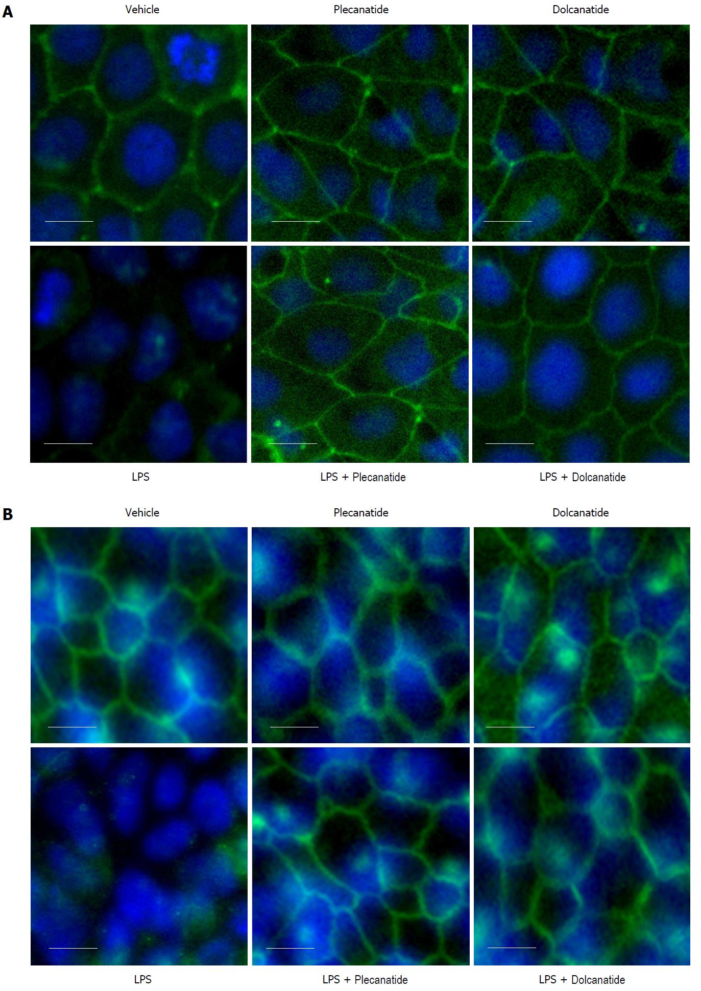
Figure 3 Effect of plecanatide and dolcanatide on localization of occludin in epithelial cells.
Caco-2 (A) and T84 (B) cell monolayers were treated with 1 μmol/L plecanatide or dolcanatide in the presence or absence of 100 μg/mL of LPS for 16 h followed by immunofluorescence imaging for occludin. Representative microscopic fields demonstrate disruption of occludin localization by LPS. Co-treatment of LPS with plecanatide or dolcanatide preserved occludin localization around the cell membrane, as was observed for vehicle treated cells. Images taken at 40 × resolution. Blue fluorescence corresponds to DAPI stained nucleus. DAPI: 4’, 6’-diamidino-2-phenylindole; LPS: lipopolysaccharide.
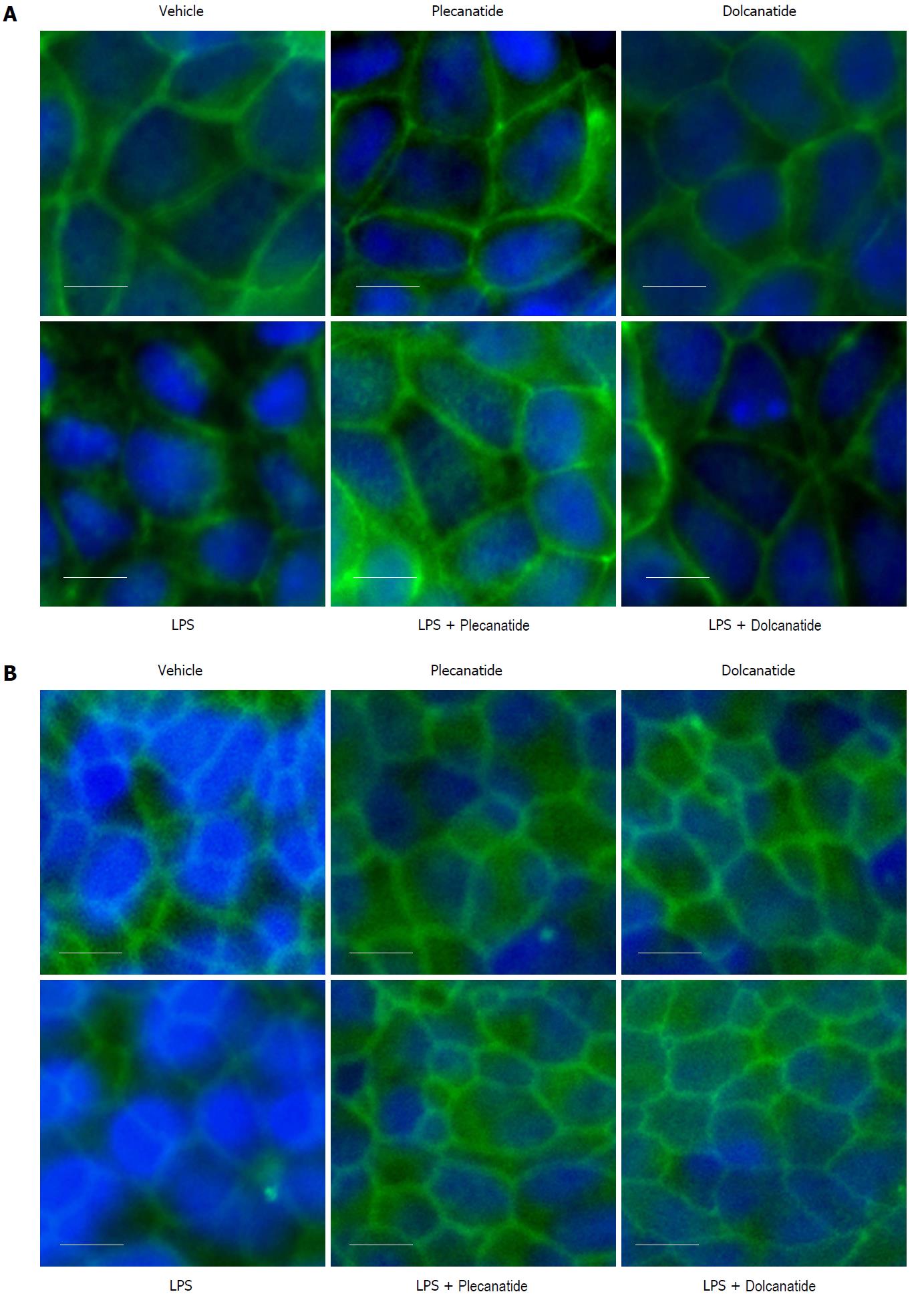
Figure 4 Effect of plecanatide and dolcanatide on localization of ZO-1 in epithelial cells Caco-2 (A) and T84 (B) cell monolayers were treated with 1 μmol/L plecanatide or dolcanatide in the presence of 100 μg/mL of LPS for 16 h followed by immunofluorescence imaging for ZO-1.
Representative microscopic fields depicted above demonstrate disruption of ZO-1 localization by LPS. In Caco-2 cells, LPS treatment appears to cause accumulation of ZO-1 in the cytoplasm. Co-treatment of LPS with plecanatide or dolcanatide preserved ZO-1 localization around the cell membrane as observed for vehicle treated cells. Images taken at 40× resolution. Blue fluorescence corresponds to DAPI stained nucleus. DAPI: 4’, 6’-Diamidino-2-phenylindole; LPS: Lipopolysaccharide; ZO-1: Zonula occludens-1.
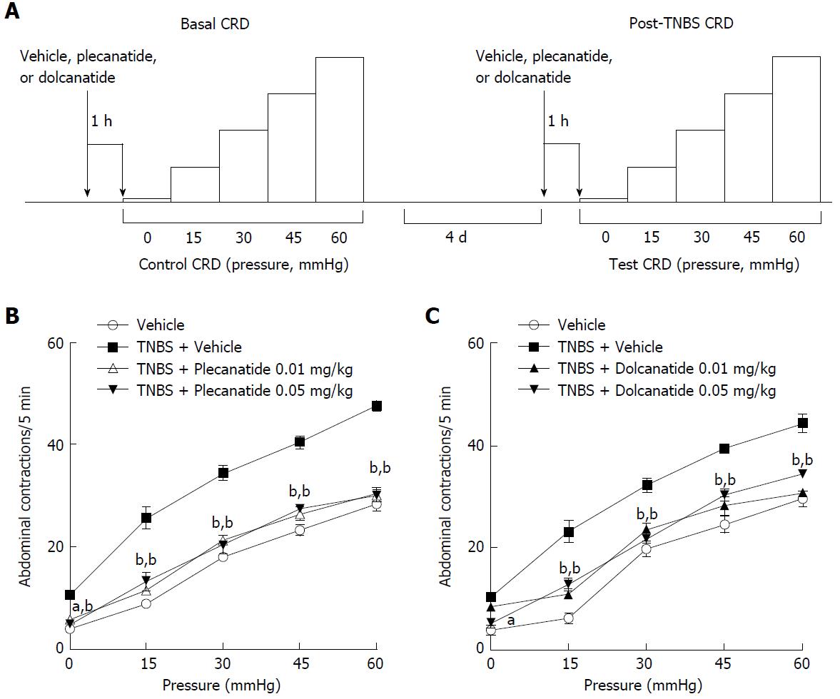
Figure 5 Design and results of the TNBS-induced visceral hypersensitivity models.
A: Schematic depicting the sequence of test sessions and treatments to evaluate visceral hypersensitivity induced by TNBS rat models. Effects of oral administration of plecanatide or dolcanatide as compared with vehicle on the increase in abdominal contractions to colorectal distention (CRD) during testing conducted four days after intrarectal administration of TNBS. Doses of 0.01 and 0.05 mg/kg of plecanatide (B) or dolcanatide (C) reduced the rate of muscular contractions toward levels observed in the vehicle group prior to TNBS administration. Data are the mean ± SEM (n = 8 rats/group). aP < 0.05, bP < 0.01 as compared to the values for the post-trinitrobenzene sulfonic acid vehicle group. SEM: Standard error of the mean; TNBS: Trinitrobenzene sulfonic acid.
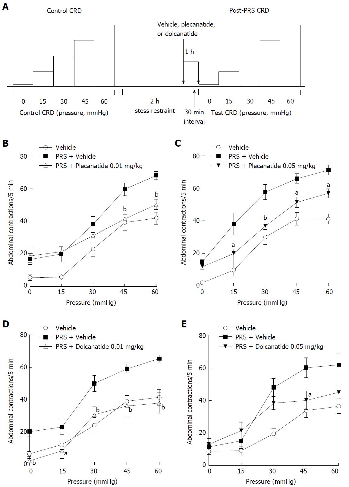
Figure 6 Design and results of the partial restraint stress-induced visceral hypersensitivity models.
A: Schematic depicting the sequence of test sessions and treatments to evaluate visceral hypersensitivity induced by PRS in rat models. Effects of oral administration of plecanatide, dolcanatide or vehicle on the increase in abdominal contractions to CRD when tested 30 min after a two h period of partial restraint. Doses of 0.01 and 0.05 mg/kg of plecanatide (B and C) or dolcanatide (D and E) 30 min before completion of the restraint session reduced the rate of muscular contractions toward the levels observed in a previous test session without exposure to partial restraint. Data are the mean ± SEM (n = 8 rats/group). aP < 0.05, bP < 0.01 as compared to the values for PRS + vehicle control. CRD: Colorectal distention; PRS: Partial restraint stress; SEM: Standard error of the mean.
- Citation: Boulete IM, Thadi A, Beaufrand C, Patwa V, Joshi A, Foss JA, Eddy EP, Eutamene H, Palejwala VA, Theodorou V, Shailubhai K. Oral treatment with plecanatide or dolcanatide attenuates visceral hypersensitivity via activation of guanylate cyclase-C in rat models. World J Gastroenterol 2018; 24(17): 1888-1900
- URL: https://www.wjgnet.com/1007-9327/full/v24/i17/1888.htm
- DOI: https://dx.doi.org/10.3748/wjg.v24.i17.1888


