Copyright
©The Author(s) 2018.
World J Gastroenterol. Apr 28, 2018; 24(16): 1803-1811
Published online Apr 28, 2018. doi: 10.3748/wjg.v24.i16.1803
Published online Apr 28, 2018. doi: 10.3748/wjg.v24.i16.1803
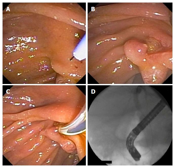
Figure 1 Schematic sequence of papillary fistulotomy.
A and B: Dissection of the major papilla; C: Sphincterotome in the bile duct; D: Radiological image.
Figure 2 Sequence of papillary fistulotomy.
A and B: Dissection of the major papilla; D: Sphincterotome in the bile duct; D: Radiological image.
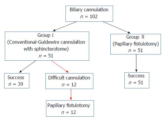
Figure 3 Flowchart showing the sequence of procedures performed in the study.
Figure 4 Amylase profile after the procedure.
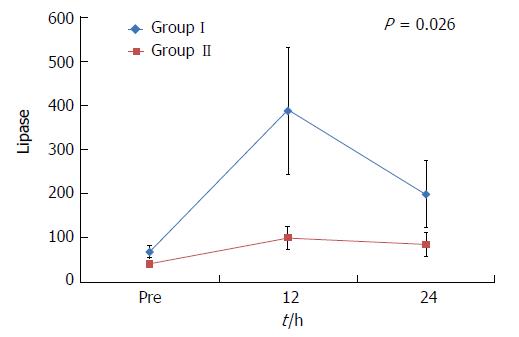
Figure 5 Lipase profile for the two groups.
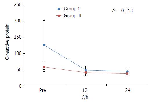
Figure 6 Evolution of C-reactive protein.
- Citation: Furuya CK, Sakai P, Marinho FRT, Otoch JP, Cheng S, Prudencio LL, de Moura EGH, Artifon ELA. Papillary fistulotomy vs conventional cannulation for endoscopic biliary access: A prospective randomized trial. World J Gastroenterol 2018; 24(16): 1803-1811
- URL: https://www.wjgnet.com/1007-9327/full/v24/i16/1803.htm
- DOI: https://dx.doi.org/10.3748/wjg.v24.i16.1803


