Copyright
©The Author(s) 2018.
World J Gastroenterol. Apr 21, 2018; 24(15): 1583-1590
Published online Apr 21, 2018. doi: 10.3748/wjg.v24.i15.1583
Published online Apr 21, 2018. doi: 10.3748/wjg.v24.i15.1583
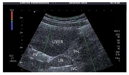
Figure 1 Enlarged perihepatic lymph nodes dorsal in the hepatoduodenal ligament between the portal vein and inferior vena cava is a typical sonographic sign of autoimmune hepatitis.
PV: Portal vein; PH: Pancreatic head; ICV: Inferior vena cava.
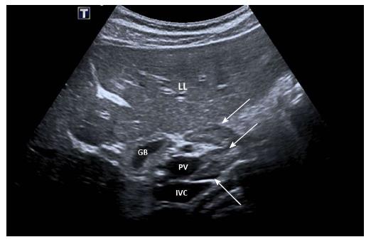
Figure 2 Enlarged perihepatic lymph nodes ventral and dorsal in the hepatoduodenal ligament between the portal vein and inferior vena cava (white arrows).
LL: Liver; GB: Galbladder; PV: Portal vein; ICV: Inferior vena cava.
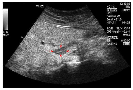
Figure 3 Enlarged perihepatic lymph nodes dorsal in the hepatoduodenal ligament is a typical sonographic sign of autoimmune hepatitis.
Contrast enhanced ultrasound shows normal lymph node architecture (in between arrows).
- Citation: Dong Y, Potthoff A, Klinger C, Barreiros AP, Pietrawski D, Dietrich CF. Ultrasound findings in autoimmune hepatitis. World J Gastroenterol 2018; 24(15): 1583-1590
- URL: https://www.wjgnet.com/1007-9327/full/v24/i15/1583.htm
- DOI: https://dx.doi.org/10.3748/wjg.v24.i15.1583


