Copyright
©The Author(s) 2018.
World J Gastroenterol. Mar 21, 2018; 24(11): 1239-1249
Published online Mar 21, 2018. doi: 10.3748/wjg.v24.i11.1239
Published online Mar 21, 2018. doi: 10.3748/wjg.v24.i11.1239
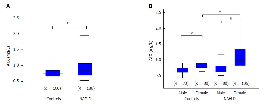
Figure 1 Comparison of autotaxin levels between controls and all patients with non-alcoholic fatty liver disease (A) and according to gender (B).
The box plot shows the interquartile range, 95% confidence interval, and median. The difference between each group was tested with the Mann Whitney U test. eP < 0.001. ATX: Autotaxin; NAFLD: Non-alcoholic fatty liver disease.
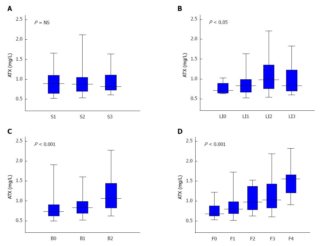
Figure 2 Relationship between autotaxin and histological grade in non-alcoholic fatty liver disease patients for steatosis (A), lobular inflammation (B), ballooning (C), and fibrosis (D).
Table 1 presents the number of subjects for each histological stage. The Kruskal-Wallis test was used for multi-group simultaneous comparisons. P values are displayed in the upper left of each graph. ATX: Autotaxin; NAFLD: Non-alcoholic fatty liver disease; NS: Not significant.
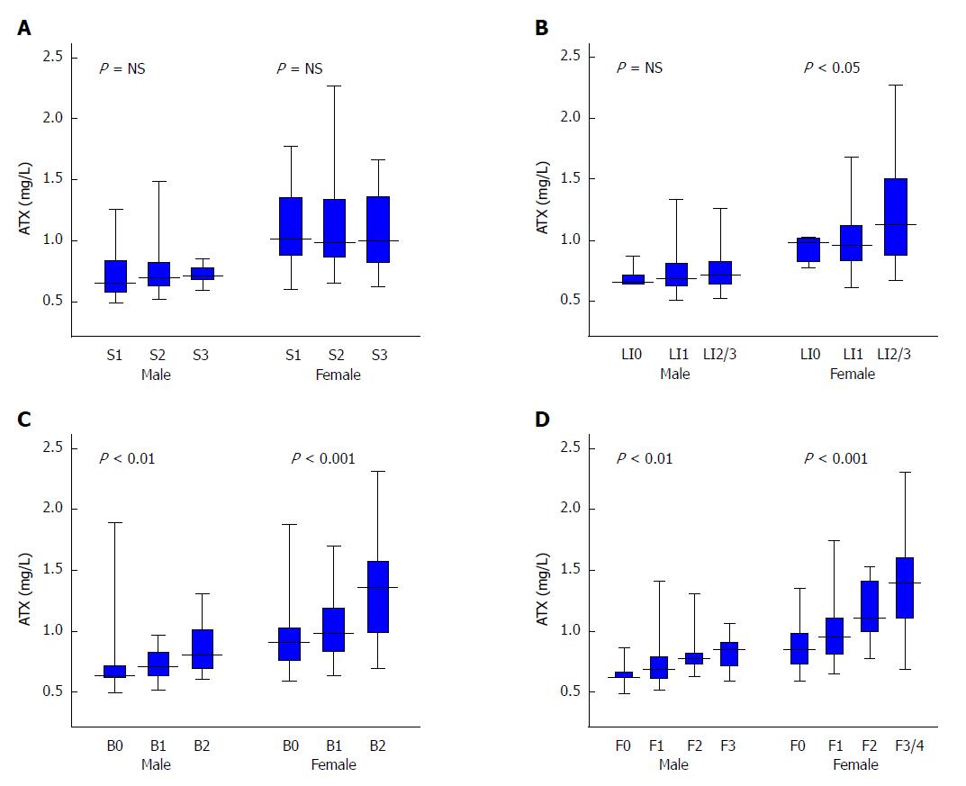
Figure 3 Relationship between autotaxin and histological grade in non-alcoholic fatty liver disease patients by gender for steatosis (A), lobular inflammation (B), ballooning (C), and fibrosis (D).
Table 1 presents the number of subjects for each histological stage. The Kruskal-Wallis test was used for multi-group simultaneous comparisons. P values are displayed in the upper left of each graph. ATX: Autotaxin; NAFLD: Non-alcoholic fatty liver disease; NS: Not significant.
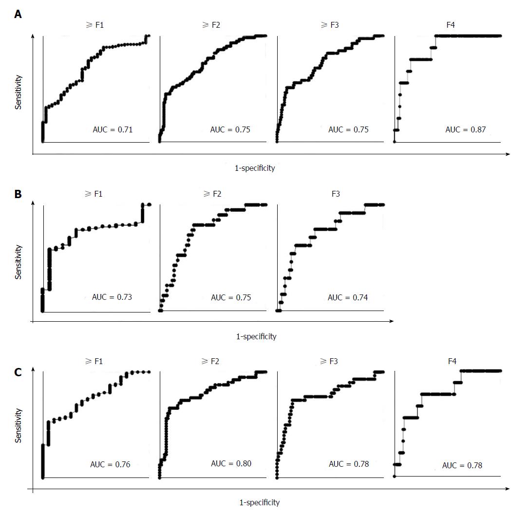
Figure 4 Receiver operating characteristic analysis of autotaxin for the estimation of the presence of fibrosis (≥ F1), significant fibrosis (≥ F2), severe fibrosis (≥ F3), and cirrhosis (F4) in all (A), male (B), and female (C) patients.
The areas under the receiver operating characteristic curve are displayed in the lower right of each graph. AUC: Receiver operating characteristic curve; F: Fibrosis.
- Citation: Fujimori N, Umemura T, Kimura T, Tanaka N, Sugiura A, Yamazaki T, Joshita S, Komatsu M, Usami Y, Sano K, Igarashi K, Matsumoto A, Tanaka E. Serum autotaxin levels are correlated with hepatic fibrosis and ballooning in patients with non-alcoholic fatty liver disease. World J Gastroenterol 2018; 24(11): 1239-1249
- URL: https://www.wjgnet.com/1007-9327/full/v24/i11/1239.htm
- DOI: https://dx.doi.org/10.3748/wjg.v24.i11.1239


