Copyright
©The Author(s) 2017.
World J Gastroenterol. Feb 14, 2017; 23(6): 1090-1097
Published online Feb 14, 2017. doi: 10.3748/wjg.v23.i6.1090
Published online Feb 14, 2017. doi: 10.3748/wjg.v23.i6.1090
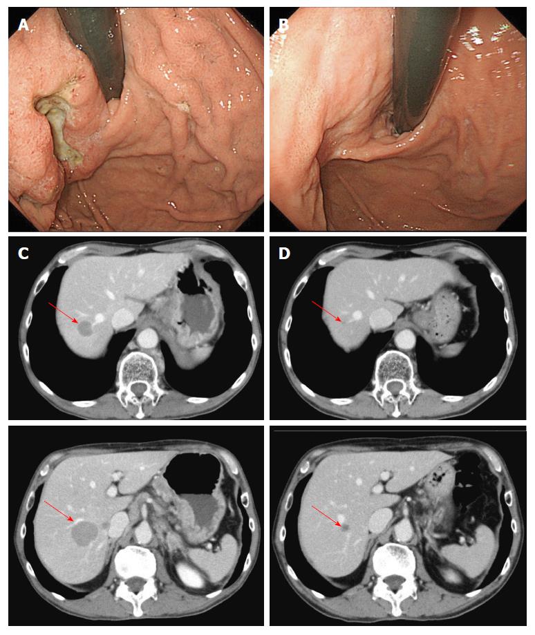
Figure 1 Case 1.
Endoscopic findings (A, B) and computed tomography images (C, D) before (A, C) and after (B, D) seven courses of modified docetaxel, cisplatin and capecitabine (DCX) (mDCX).
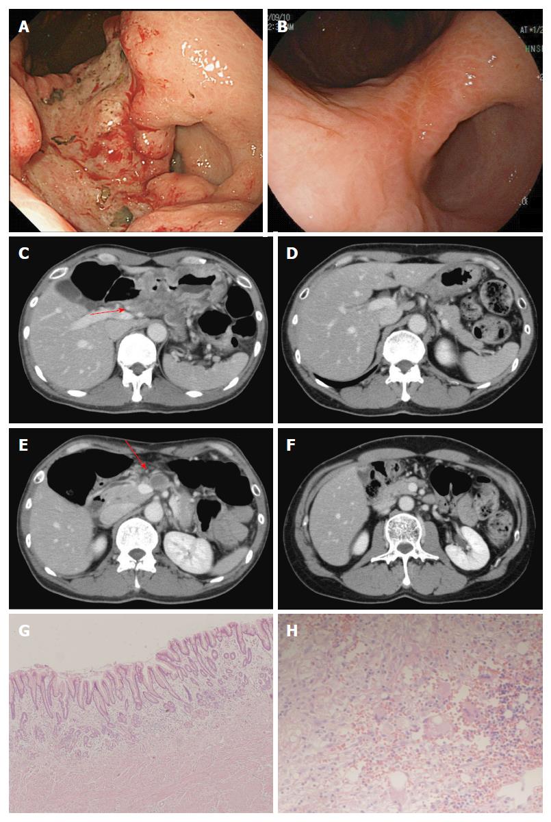
Figure 2 Case 2.
Endoscopic findings (A, B) and computed tomography images (C, D, E, F) before treatment (A, C, E) and after (B, D, F) five courses of modified docetaxel, cisplatin and capecitabine (DCX) (mDCX). The primary lesion (A, B), swollen lymph nodes along the common hepatic artery (#8) (C, D) and lymph nodes along the superior mesenteric artery (#14a) (E, F) markedly shrank. Microscopic findings for the resected specimens of the primary lesion (E, magnification × 100) and lymph nodes (F, magnification × 400) revealed no residual tumor.
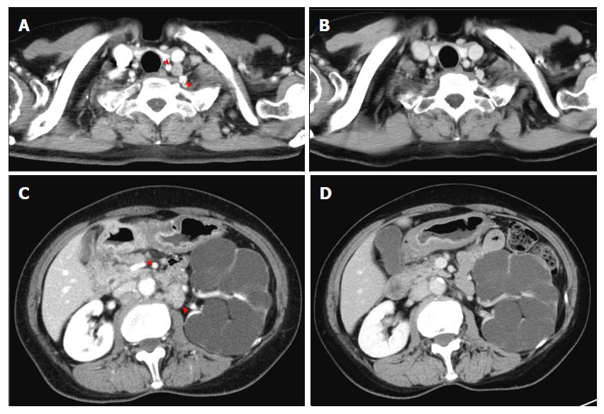
Figure 3 Case 8.
Computed tomography images before (A, C) and after (B, D) four courses of modified docetaxel, cisplatin and capecitabine (DCX) (mDCX). Left supraclavicular nodes (A, B) and para-aortic nodes (C, D) became undetectable.
- Citation: Maeda O, Matsuoka A, Miyahara R, Funasaka K, Hirooka Y, Fukaya M, Nagino M, Kodera Y, Goto H, Ando Y. Modified docetaxel, cisplatin and capecitabine for stage IV gastric cancer in Japanese patients: A feasibility study. World J Gastroenterol 2017; 23(6): 1090-1097
- URL: https://www.wjgnet.com/1007-9327/full/v23/i6/1090.htm
- DOI: https://dx.doi.org/10.3748/wjg.v23.i6.1090


