Copyright
©The Author(s) 2017.
World J Gastroenterol. Dec 28, 2017; 23(48): 8660-8665
Published online Dec 28, 2017. doi: 10.3748/wjg.v23.i48.8660
Published online Dec 28, 2017. doi: 10.3748/wjg.v23.i48.8660
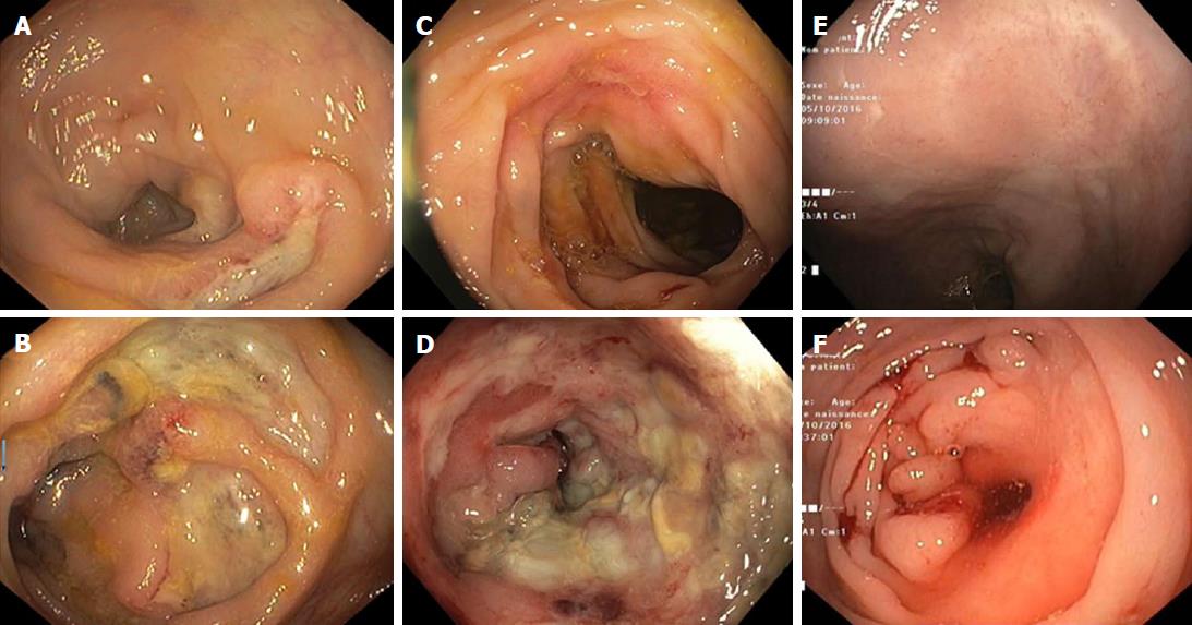
Figure 1 Endoscopic finding before and after withdrawal of belatacept.
The first colonoscopy showed the presence of disseminated ulcers on the colonic mucosa (A) with more severe lesions at the left colonic flexure (B). The patient was still on mycophenolate mofetil and belatacept. After mycophenolate withdrawal and continuation of belatacept, the second colonoscopy showed the persistence of disseminated ulcerations (C) and a worsening of lesions at the left colonic flexure with appearance of an inflammatory ulcerated stricture (D). Five months after belatacept withdrawal, follow up colonoscopy showed complete healing of disseminated ulcerations in the left colon (transverse and right colon were not visualized due to the stricture, (E) and healing of the left colonic flexure with persistence of a non-inflammatory stenosis (F) which did not allow passage of the colonoscope.
Figure 2 Histologic findings of belatacept-induced colitis.
A: Histologic examination of colonic biopsies showed acute colitis with ulcerations, crypt abscesses, lymphocytes and neutrophil polymorphonuclear leukocyte infiltration. Neither crypt dystrophy nor granuloma was found; B: After belatacept withdrawal, colonic biopsies showed complete healing of the mucosa with no signs of chronic mucosal inflammation.
- Citation: Bozon A, Jeantet G, Rivière B, Funakoshi N, Dufour G, Combes R, Valats JC, Delmas S, Serre JE, Bismuth M, Ramos J, Le Quintrec M, Blanc P, Pineton de Chambrun G. Stricturing Crohn’s disease-like colitis in a patient treated with belatacept. World J Gastroenterol 2017; 23(48): 8660-8665
- URL: https://www.wjgnet.com/1007-9327/full/v23/i48/8660.htm
- DOI: https://dx.doi.org/10.3748/wjg.v23.i48.8660


