Copyright
©The Author(s) 2017.
World J Gastroenterol. Dec 28, 2017; 23(48): 8553-8561
Published online Dec 28, 2017. doi: 10.3748/wjg.v23.i48.8553
Published online Dec 28, 2017. doi: 10.3748/wjg.v23.i48.8553
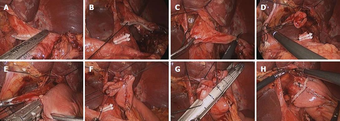
Figure 1 Forming an esophagojejunostomy.
A: Nearly two-thirds of the esophagus diameter is transected 2 cm above the gastroesophageal junction using an endoscopic linear stapler; B: The first intracorporeal suture is made at the end of the staple line of the esophageal stump; C: The unstapled esophagus is transected with laparoscopic scissors after the remnant stomach has been clipped with manual titanium clips to avoid spillage of cancer cells; D: The second and third intracorporeal sutures are made at the esophagostomy site of the esophageal stump; E: To create an esophagojejunostomy, an endoscopic linear stapler is inserted by the operator between the esophagostomy and enterostomy of the jejunum. At this time the first assistant retracts the first thread towards the operator’s direction inside the abdominal cavity, and the second assistant retracts the second thread through the right lower trocar from the outside of the abdomen; F: After an esophagojejunostomy has been constructed, the entry hole is held with tress suturing to approximate the tissue; G: The remnant entry hole is closed by the operator with an endoscopic linear stapler; H: An esophagojejunal anastomosis after completion of the reconstruction.
- Citation: Gong CS, Kim BS, Kim HS. Comparison of totally laparoscopic total gastrectomy using an endoscopic linear stapler with laparoscopic-assisted total gastrectomy using a circular stapler in patients with gastric cancer: A single-center experience. World J Gastroenterol 2017; 23(48): 8553-8561
- URL: https://www.wjgnet.com/1007-9327/full/v23/i48/8553.htm
- DOI: https://dx.doi.org/10.3748/wjg.v23.i48.8553


