Copyright
©The Author(s) 2017.
World J Gastroenterol. Dec 28, 2017; 23(48): 8526-8532
Published online Dec 28, 2017. doi: 10.3748/wjg.v23.i48.8526
Published online Dec 28, 2017. doi: 10.3748/wjg.v23.i48.8526
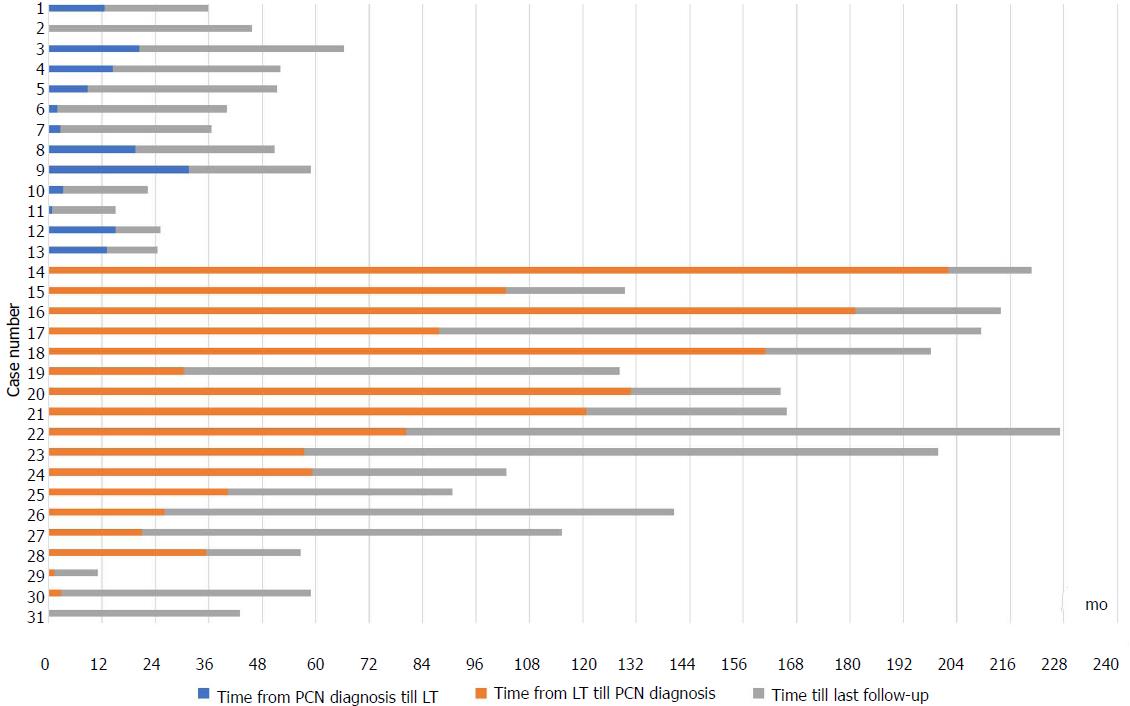
Figure 1 Time points for pancreatic cystic neoplasms diagnosis, liver transplantation and follow-up in the 31 patients with pancreatic cystic neoplasms.
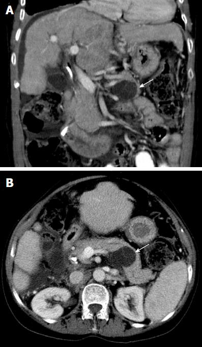
Figure 2 Coronal (A) and axial (B) computed tomography images in portal venous phase demonstrate a well-defined low density lesion in the body/tail of the pancreas measuring 40 mm in maximum transverse dimensions (white arrows).
Thin septa are demonstrated within the cyst.
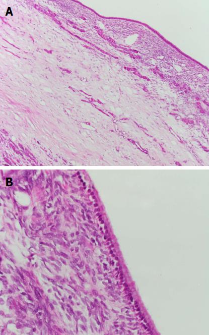
Figure 3 Lower power 100 × magnification view (A) and higher power 400 × magnification view (B) of a mucinous cystic neoplasm with low grade dysplasia.
The cyst is lined by columnar mucinous epithelium with underlying ovarian-type stroma (hematoxylin-eosin stain).
- Citation: Liu K, Joshi V, van Camp L, Yang QW, Baars JE, Strasser SI, McCaughan GW, Majumdar A, Saxena P, Kaffes AJ. Prevalence and outcomes of pancreatic cystic neoplasms in liver transplant recipients. World J Gastroenterol 2017; 23(48): 8526-8532
- URL: https://www.wjgnet.com/1007-9327/full/v23/i48/8526.htm
- DOI: https://dx.doi.org/10.3748/wjg.v23.i48.8526


