Copyright
©The Author(s) 2017.
World J Gastroenterol. Dec 7, 2017; 23(45): 8073-8081
Published online Dec 7, 2017. doi: 10.3748/wjg.v23.i45.8073
Published online Dec 7, 2017. doi: 10.3748/wjg.v23.i45.8073
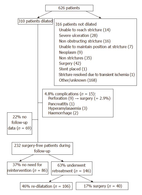
Figure 1 Study results.
This flowchart summarizes the study results with the outcomes of the patients that were dilated.
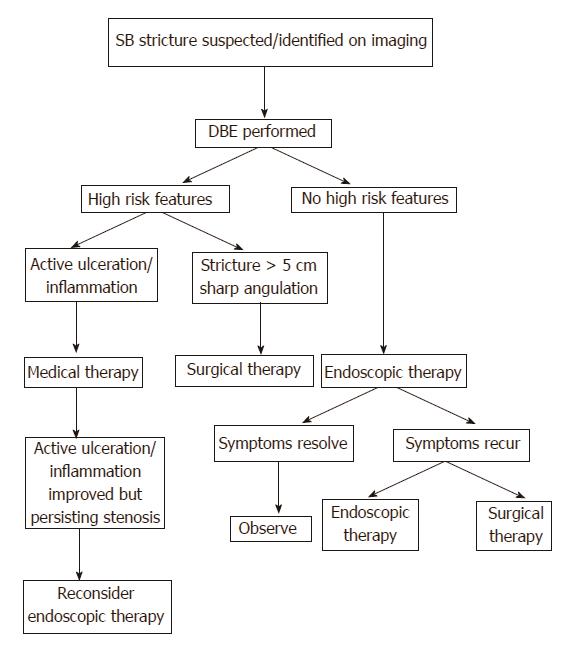
Figure 2 Suggested approach to small bowel.
This algorithm proposes a standardized approach to small bowel strictures, taking into account the known risk factors previously demonstrated in literature.
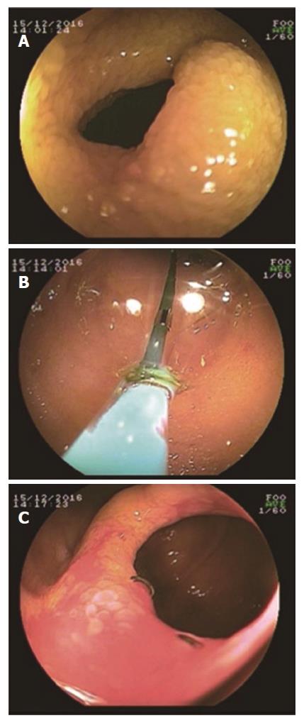
Figure 3 Double-balloon enteroscopy -assisted balloon dilatation.
An example of a successful double-balloon enteroscopy (DBE)-assisted balloon dilatation is presented. A: shows the endoscopic image of a benign small bowel stricture in one of our patients. This patient was known with Crohn’s disease and had had prior small bowel surgery. She presented with obstructive symptoms and a fibrotic stricture at the side of the anastomosis. B: The stricture was dilated with DBE-assisted balloon dilation; C: Shows the anastomotic stricture after successful dilatation. This picture reveals the surgical staples at the anastomosis and there were no signs of active Crohn’s disease.
- Citation: Baars JE, Theyventhiran R, Aepli P, Saxena P, Kaffes AJ. Double-balloon enteroscopy-assisted dilatation avoids surgery for small bowel strictures: A systematic review. World J Gastroenterol 2017; 23(45): 8073-8081
- URL: https://www.wjgnet.com/1007-9327/full/v23/i45/8073.htm
- DOI: https://dx.doi.org/10.3748/wjg.v23.i45.8073


