Copyright
©The Author(s) 2017.
World J Gastroenterol. Dec 7, 2017; 23(45): 7945-7951
Published online Dec 7, 2017. doi: 10.3748/wjg.v23.i45.7945
Published online Dec 7, 2017. doi: 10.3748/wjg.v23.i45.7945
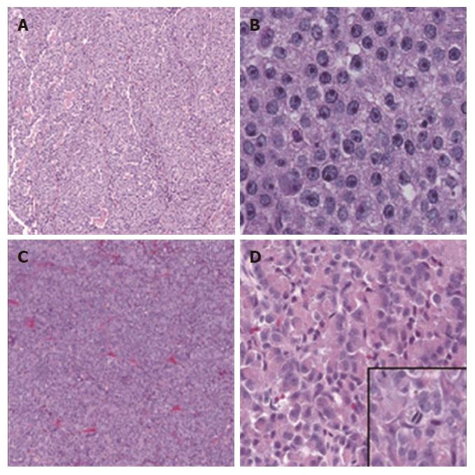
Figure 1 Different histological forms of pancreatic acinar cell carcinomas.
A: A case of PACC displaying nested to glandular growth patterns (HE 40 ×); B: Higher magnification of the same tumor in Panel A showing monotonous cells with eosinophilic/granular cytoplasm with well-defined cell borders and uniform nuclei with minimal atypia and prominent nucleoli (HE 200 ×); C: Tumor from a different patient showing a predominantly sheet-like growth with no distinct pattern (HE 40 ×). D: Higher magnification of the same tumor in C showing uniform cells with eosinophilic granular cytoplasm (prominent zymogen granules) with minimal pleomorphism (HE 200 ×). Inset: mitotic figures were identified throughout the tumor (HE 400 ×).
- Citation: Al-Hader A, Al-Rohil RN, Han H, Von Hoff D. Pancreatic acinar cell carcinoma: A review on molecular profiling of patient tumors. World J Gastroenterol 2017; 23(45): 7945-7951
- URL: https://www.wjgnet.com/1007-9327/full/v23/i45/7945.htm
- DOI: https://dx.doi.org/10.3748/wjg.v23.i45.7945


