Copyright
©The Author(s) 2017.
World J Gastroenterol. Nov 14, 2017; 23(42): 7572-7583
Published online Nov 14, 2017. doi: 10.3748/wjg.v23.i42.7572
Published online Nov 14, 2017. doi: 10.3748/wjg.v23.i42.7572
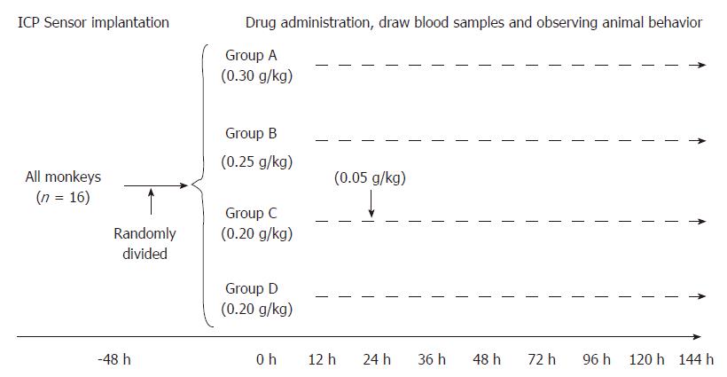
Figure 1 Study design.
All monkeys were randomly divided into four groups after ICP sensor implantation; the interval of D-gal administration to group C was 24 h. ICP: Intracranial pressure.
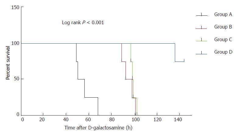
Figure 2 Survival times of monkeys in different study groups.
Group A vs group B: P = 0.007; group A vs group C: P = 0.007; Group A vs group D: P < 0.001; Group B vs group C: P = 0.375.
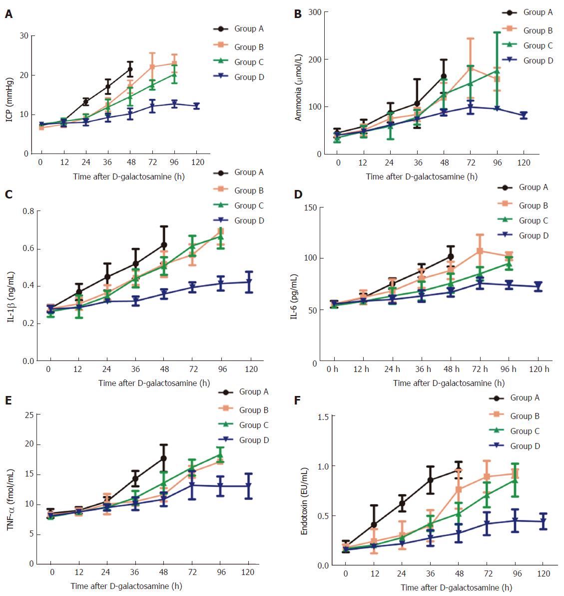
Figure 3 Changes of intracranial pressure, ammonia, inflammation markers and endotoxin at different time points in each group.
All data points are mean ± SD, n = 4. ICP: Intracranial pressure; Amm: Ammonia; IL-1β: Interleukin-1β; IL-6: Interleukin-6; TNF-α: Tumor necrosis factor-α.
Figure 4 Changes of biochemical indices at different time points in each group.
All data points are mean ± SD, n = 4. ALT: Alanine aminotransferase; AST: Aspartate aminotransferase; TBIL: Total bilirubin; PT: Prothrombin time; ALB: Albumin; LDH: Lactic dehydrogenase; CK: Creatine kinase; BUN: Blood urea nitrogen; Cr: Creaninine.
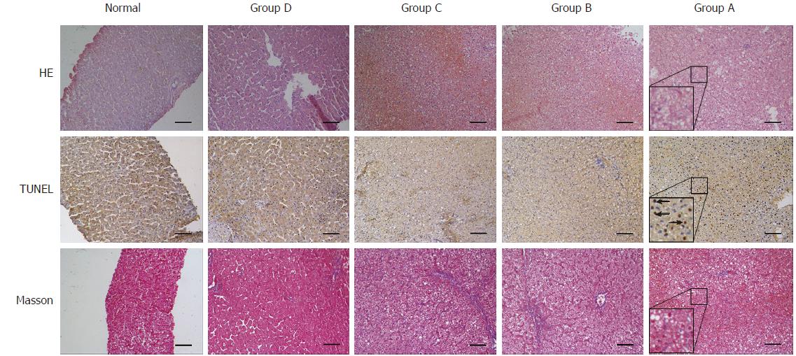
Figure 5 H&E staining, Tunel and Masson assays of post-mortem liver specimens from different groups.
H&E: Hematoxylin-eosin staining; TUNEL: Terminal -deoxynucleotidyl transferase mediated nick end labeling; Arrows: Apoptotic bodies. Lower left corner detail: enlarged scale for group A (× 100 magnification, 200 μm scale bars).
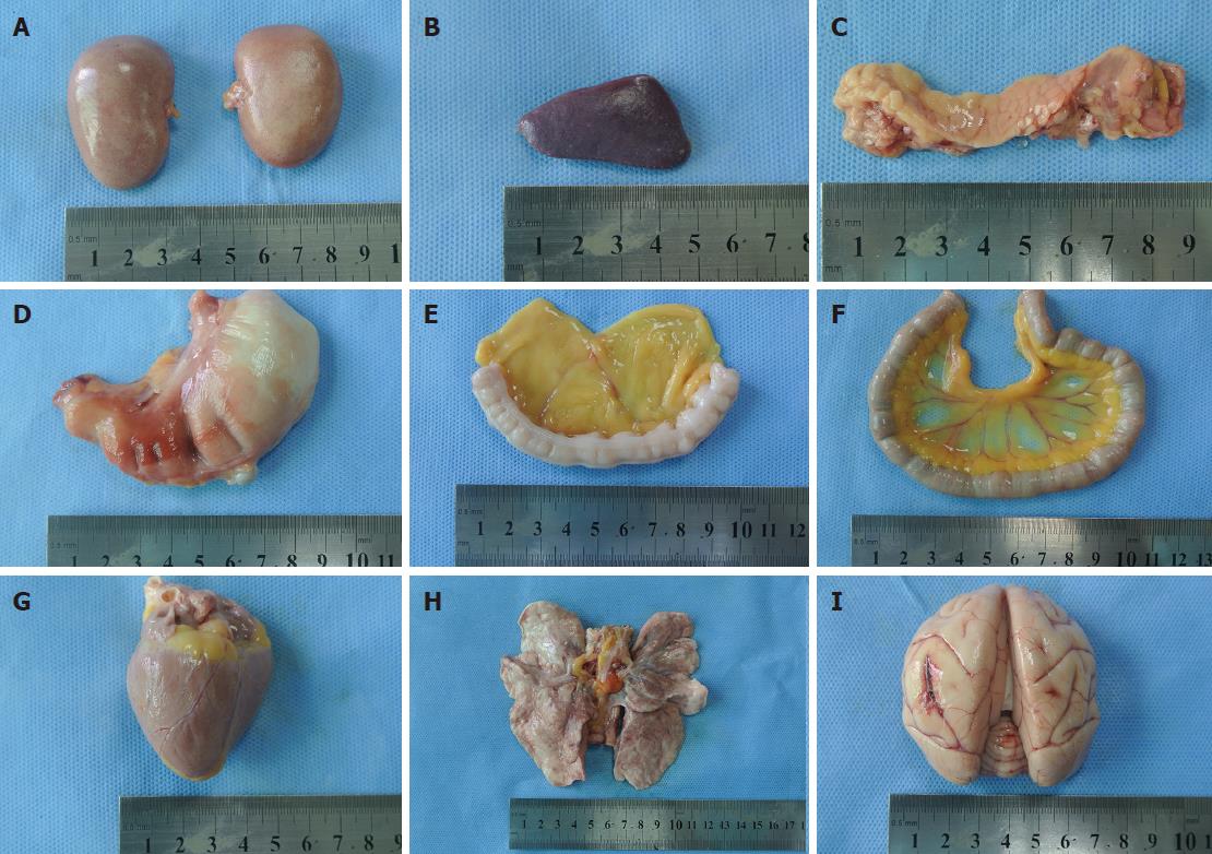
Figure 6 Gross specimens of other organs post-mortem (in group C).
A: Renal; B: Spleen; C: Pancreas; D: Stomach; E: Large intestine; F: Small intestine;G: Heart; H: Lung; I: Brain.
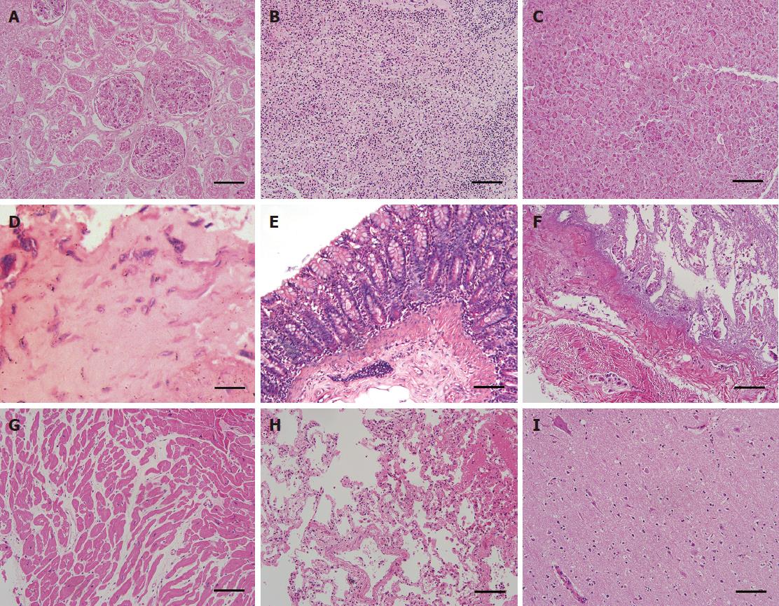
Figure 7 HE staining of other organs post-mortem (in group C).
A: The renal tissue profile was clear, and the glomerular capillaries and renal interstitial blood vessels were slightly dilated and congested; B: The splenic sinusoids were mildly to moderately expanded with a large number of red blood cells; C: Pancreas, no abnormalities; D: Stomach, no abnormalities; E: Large intestine, no abnormalities; F: Small intestine, no abnormalities; G: Heart, no abnormalities; H: Lung, the bronchial and alveolar structures of pulmonary tissues were complete, and the interstitial capillaries were diffusely expanded and congested with few red blood cells; I: Brain, the nerve cells were diffusely enlarged with mild degenerative changes (× 100 magnification, 200 μm scale bars).
- Citation: Feng L, Cai L, He GL, Weng J, Li Y, Pan MX, Jiang ZS, Peng Q, Gao Y. Novel D-galactosamine-induced cynomolgus monkey model of acute liver failure. World J Gastroenterol 2017; 23(42): 7572-7583
- URL: https://www.wjgnet.com/1007-9327/full/v23/i42/7572.htm
- DOI: https://dx.doi.org/10.3748/wjg.v23.i42.7572


