Copyright
©The Author(s) 2017.
World J Gastroenterol. Jan 28, 2017; 23(4): 730-734
Published online Jan 28, 2017. doi: 10.3748/wjg.v23.i4.730
Published online Jan 28, 2017. doi: 10.3748/wjg.v23.i4.730
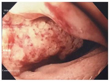
Figure 1 Gastroscopy shows a circumferential vegetating mass with a villous appearance on the second portion of the duodenum.
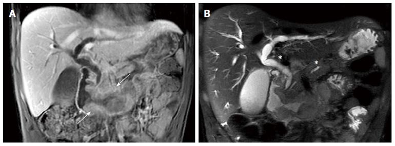
Figure 2 Magnetic resonance imaging.
A: MRI, coronal T1-weighted MR image with contrast showing the duodenal tissue thickening spreading from the second duodenum proximal part up to the duodenojejunal flexure (white arrow); B: MRI, coronal T2-weighted MR image showing the pancreatic duct and bile duct upstream swelling (white asterisks) without any secondary hepatic lesion. MRI: Magnetic resonance imaging.
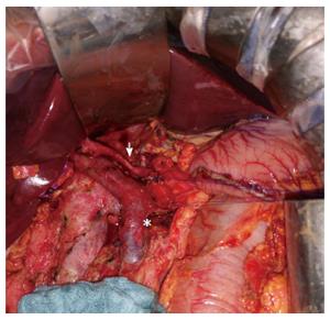
Figure 3 Intraoperative view of the pancreaticoduodenectomy showing lymph node dissection with excision of all cellular adipose tissues of the hepatic pedicle from the common hepatic artery (white arrow) and of the retroportal lamina (white asterisk).
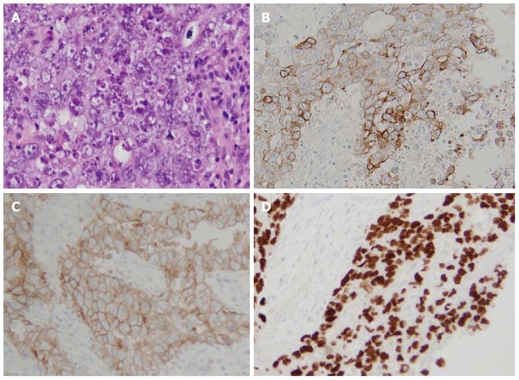
Figure 4 Histological findings of the operative specimen showing an embryonal carcinoma: pleomorphic cell proliferation with marked cytonuclear atypia, granular or clear cytoplasm, arranged in nests or solid pattern, associated with a fibrous stroma and an abundant lymphocytic inflammatory infiltrate (A).
Specimen cells expressing cytokeratin 7 (B), CD30 (C) and SALL4 (D) are evidenced in immunohistochemical staining. A: HE × 400; B: Cytokeratin 7 × 200; C: CD30 × 200; D: SALL4 × 200.
- Citation: Barbieux J, Memeo R, De Blasi V, Suciu S, Faucher V, Averous G, Roy C, Marescaux J, Mutter D, Pessaux P. Real case of primitive embryonal duodenal carcinoma in a young man. World J Gastroenterol 2017; 23(4): 730-734
- URL: https://www.wjgnet.com/1007-9327/full/v23/i4/730.htm
- DOI: https://dx.doi.org/10.3748/wjg.v23.i4.730


