Copyright
©The Author(s) 2017.
World J Gastroenterol. Oct 21, 2017; 23(39): 7185-7190
Published online Oct 21, 2017. doi: 10.3748/wjg.v23.i39.7185
Published online Oct 21, 2017. doi: 10.3748/wjg.v23.i39.7185
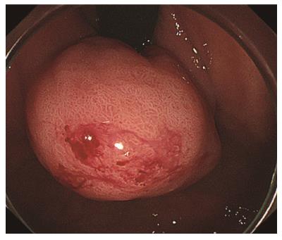
Figure 1 Endoscopic findings of gastric submucosal tumor.
A gastric submucosal tumor (30 mm in diameter) is shown in the fornix of the stomach.
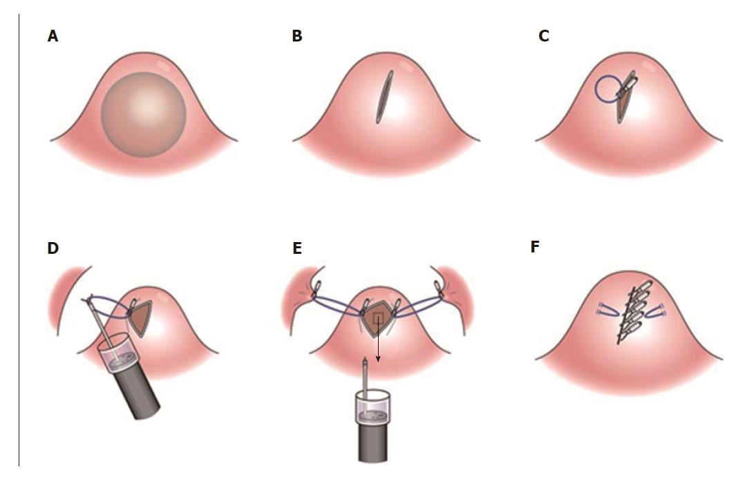
Figure 2 Oval mucosal opening bloc biopsy after incision and widening by ring thread traction.
A: A gastric submucosal tumor (SMT) (30 mm in diameter) is shown in the fornix of the stomach; B: A 5-10 mm incision on the top of SMT was made; C: After a 5-mm ring-shaped thread was delivered by grasping forceps; D: Second clip was hooked the ring-shaped thread and moved to be tied up the left gastric wall; E: The same procedures were performed on the right side of the incision mucosa and made a straight incision like an oval-shaped incision; F: After both sides of the ring threads were detached, the opened mucosa was closed by hemoclips to restore it back to the original mucosa.
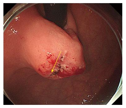
Figure 3 Incision at the top of the submucosal tumor.
As endoscopic ultrasound sound fine needle aspiration and submucosal tunneling bloc biopsy were impossible due to the tumor’s location, a 5-10 mm incision on the top of submucosal tumor was made (yellow arrow).
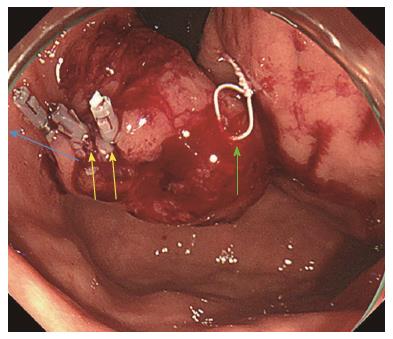
Figure 4 Ring- shaped thread counter traction.
After clipping the 5-mm ring-shaped thread on the left side mucosa of the incision edge (yellow arrows), the other side of this ring thread was hooked and pulled to the posterior wall of the stomach (blue arrow). A 2nd white ring thread was placed on the other side of the incision edge (green arrow).
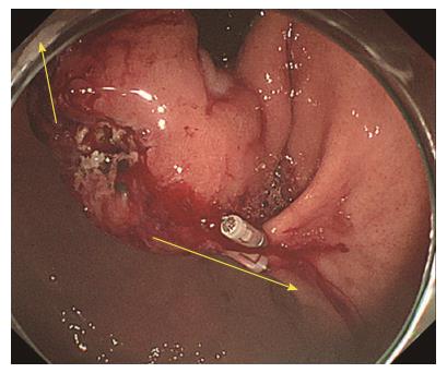
Figure 5 Oval mucosal opening after incision and widening by ring thread traction.
The same procedures were performed on both sides of the incision mucosa with a straight incision to an oval shaped incision (yellow arrows).
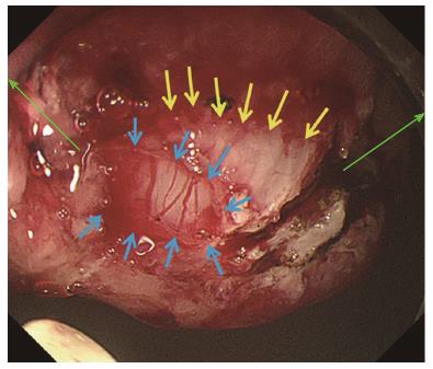
Figure 6 Direct view of capsule and abundant vessels of gastrointestinal stromal tumors.
With more insufflation, both ring threads expanded the oval incision to a round shaped incision (green arrows) from which the tumor capsule was clearly recognized. An approximately 7-mm cut of the tumor capsule (yellow arrows) by Dual knife made it possible to confirm the tumor (blue arrows) with abundant tumor vessels.
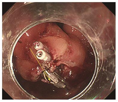
Figure 7 Reversible mucosa closure by hemoclips.
After both sides of the ring threads were detached, the opened mucosa was closed by hemoclips to restore it back to the original mucosa (yellow arrow).
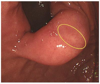
Figure 8 A mucosal incision six week after oval mucosal opening bloc biopsy.
An endoscopic image revealed that straight incision on the top of the submucosal tumor was completely scarred and closed (yellow ring) when laparoscopy and endoscopy cooperative surgery was performed six week after oval mucosal opening bloc biopsy.
- Citation: Mori H, Kobara H, Guan Y, Goda Y, Kobayashi N, Nishiyama N, Masaki T. Oval mucosal opening bloc biopsy after incision and widening by ring thread traction for submucosal tumor. World J Gastroenterol 2017; 23(39): 7185-7190
- URL: https://www.wjgnet.com/1007-9327/full/v23/i39/7185.htm
- DOI: https://dx.doi.org/10.3748/wjg.v23.i39.7185


