Copyright
©The Author(s) 2017.
World J Gastroenterol. Oct 21, 2017; 23(39): 7150-7159
Published online Oct 21, 2017. doi: 10.3748/wjg.v23.i39.7150
Published online Oct 21, 2017. doi: 10.3748/wjg.v23.i39.7150
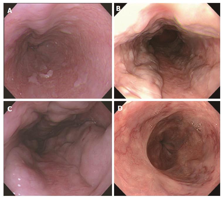
Figure 1 Classification of esophageal varices.
A: F1 varices, small and straight; B: F2 varices, nodular; C: F3 varices, large or coiled; D: post-treatment varices.
Figure 2 Portal hypertensive gastropathy lesions exhibiting a snake-skin (mosaic) pattern in their background mucosa.
A: Grade 1, erythematous flecks or maculae; B: Grade 2, red spots or diffuse redness; C: Grade 3, intramucosal or luminal hemorrhage.
Figure 3 Columnar-lined esophagus and hiatal hernia.
A: CLE 10-29 mm; B: CLE 5-9 mm; C: CLE 0-4 mm and hiatal hernia ≥ 10 mm. The endoscopic esophagogastric junction was defined as the lower limit of the palisade longitudinal vessels (arrows). The axial length of a hiatal hernia was defined as the distance between the esophagogastric junction and the hiatus represented by the diaphragmatic pinch. CLE: Columnar-lined esophagus.
- Citation: Yokoyama A, Hirata K, Nakamura R, Omori T, Mizukami T, Aida J, Maruyama K, Yokoyama T. Presence of columnar-lined esophagus is negatively associated with the presence of esophageal varices in Japanese alcoholic men. World J Gastroenterol 2017; 23(39): 7150-7159
- URL: https://www.wjgnet.com/1007-9327/full/v23/i39/7150.htm
- DOI: https://dx.doi.org/10.3748/wjg.v23.i39.7150


