Copyright
©The Author(s) 2017.
World J Gastroenterol. Oct 14, 2017; 23(38): 6931-6941
Published online Oct 14, 2017. doi: 10.3748/wjg.v23.i38.6931
Published online Oct 14, 2017. doi: 10.3748/wjg.v23.i38.6931
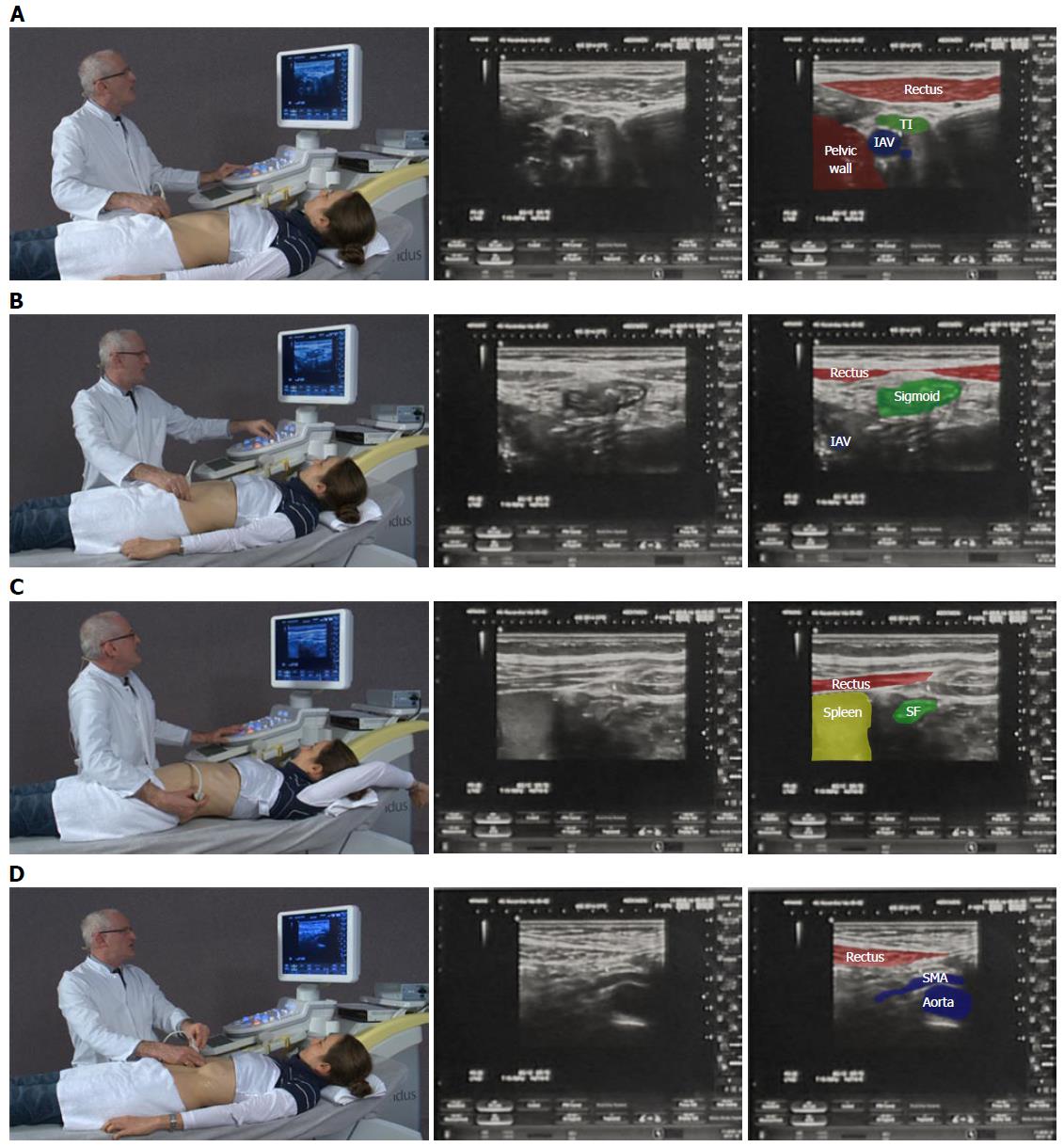
Figure 1 A systematic approach in examining the whole intestine.
A: Examination begins in a relaxed ventral position; B: Beginning medial to the right anterior superior iliac spine, the iliacal vessels (IAV) are identified and the first bowel loop crossing medial-to-lateral is the terminal ileum (TI). The same technique on the right identifies the sigmoid colon; C: Elevating the arm spreads the rib spaces to improve visualisation of the splenic flexure (SF); D: Gentle pressure as the patient breaths out improves visualization of the mesentery and superior mesenteric artery (SMA) to exclude lymphadenopathy. The videos can be accessed via the efsumb website [http://www.efsumb.org/education/cfd-videos001.asp].
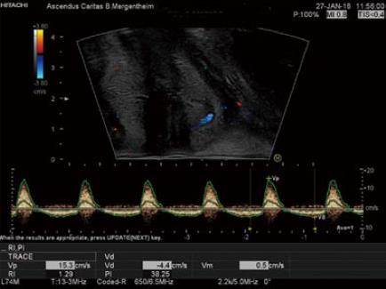
Figure 2 Example on the use of color doppler imaging and continous duplex scanning.
Perineal ultrasound showing the hemorrhoidal pleaxus using color doppler imaging and continous duplex scanning with the typical spectrum of the hemorrhoids.
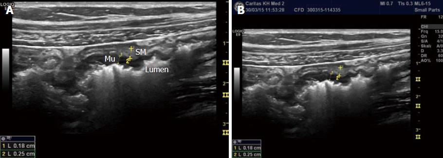
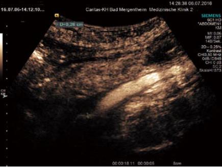
Figure 3 Measurement of the bowel wall.
The measurements are best taken ventrally since posterior artefacts occur (A) and the measurements (B) are not reliable. Mu: Mucosa; SM: Submucosa.
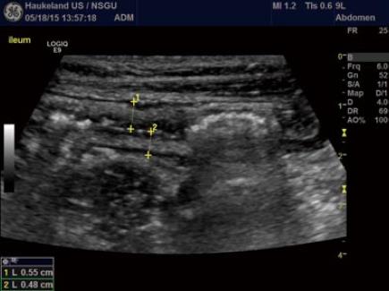
Figure 4 Measurement of the bowel wall.
In a patient with Crohn’s disease of the small intestine, ultrasound was applied to evaluate disease extension and wall thickness. B-mode image shows moderate wall thickening in the ileum with well-preserved layer structure. Be aware the marked thickening of the submucosal layer in white, often seen in IBD. The crosses mark the wall thickness in the anterior and posterior wall denoting a slight difference in thickening.
Figure 5 Typical complications in Crohn’s disease, fistula.
Typical ultrasound findings in Crohn’s disease include transmural inflammation, fistula and abscess formation. A-C: The typical sign of fistula is hypoechoic transmural inflammation with (moving) air bubbles outside of the bowel lumen. The air bubbles are best visualised using a real-time examination or video. Here we demonstrate single images of a video to demonstrate the changes within one second.
Figure 6 Typical complications in Crohn’s disease, abscess.
Typical ultrasound findings in Crohn’s disease include transmural inflammation, fistula and abscess formation. Contrast enhanced ultrasound allows to better delineate larger (A) and smaller abscess formation (B) not clearly suspected using B mode ultrasound.
Figure 7 Complications of inflammatory bowel disease.
Thrombosis of the superior mesenteric vein. Partial recanalisation is shown by the markers.
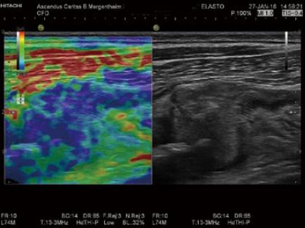
Figure 8 The evaluation of bowel wall stiffness.
Elastography is helpful to determine stiff tissue as shown in this patient with colorectal carcinoma and infiltration of the abdominal wall.
- Citation: Atkinson NSS, Bryant RV, Dong Y, Maaser C, Kucharzik T, Maconi G, Asthana AK, Blaivas M, Goudie A, Gilja OH, Nuernberg D, Schreiber-Dietrich D, Dietrich CF. How to perform gastrointestinal ultrasound: Anatomy and normal findings. World J Gastroenterol 2017; 23(38): 6931-6941
- URL: https://www.wjgnet.com/1007-9327/full/v23/i38/6931.htm
- DOI: https://dx.doi.org/10.3748/wjg.v23.i38.6931


