Copyright
©The Author(s) 2017.
World J Gastroenterol. Oct 7, 2017; 23(37): 6750-6776
Published online Oct 7, 2017. doi: 10.3748/wjg.v23.i37.6750
Published online Oct 7, 2017. doi: 10.3748/wjg.v23.i37.6750
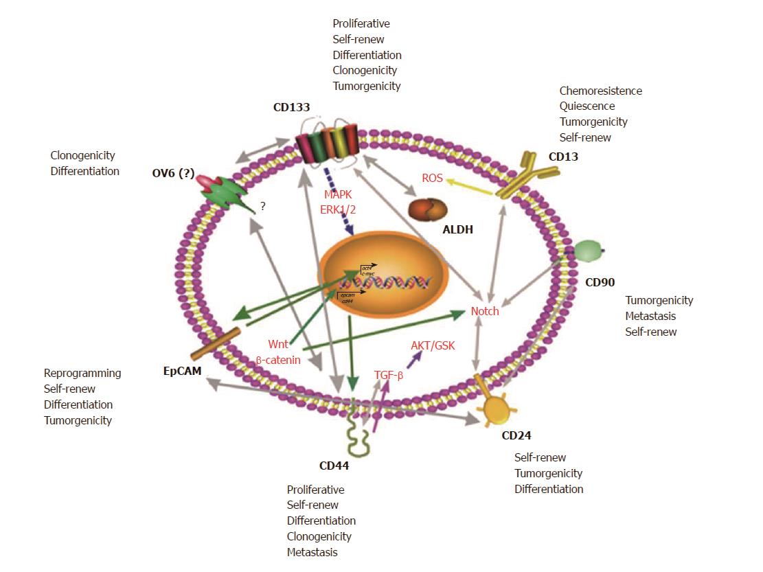
Figure 1 One marker is not enough for designate one cell as cancer stem cell.
Some markers are reported as representative of HCSCs (CD133, CD13, CD90, CD24, CD44, EpCAM, OV6 and activity of the ALDH family) nevertheless there are main signaling pathways dictating the stemness state. OV6 structure and function is still unknown. Properties or characteristics related of every HCSC marker are listed besides each marker (self-renew, chemoresistence, quiescence, tumorgenicity, etc). Gray lines with double arrows indicate correlation in expression (i.e. Notch and CD90, CD133 and CD13 are overexpressed in the same CSCs population). Straight lines indicate that the molecule or pathway affects directly another molecule or pathway (i.e. CD44 affects directly TGF-β pathway, and TGF-β pathway affects directly AKT/GSK pathway). Doted line indicates that it affects indirectly the molecule or pathway (i.e., CD133 expression affects IL-8/CXCL1 via MAPK/ERK indirect activation).
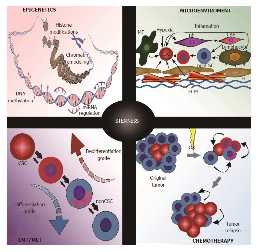
Figure 2 Stimuli that trigger the gain of stemness of non-cancer stem cells in hepatocellular carcinoma.
The diagram includes the conditions that are reported to induce stemness: epigenetics (histone modifications, DNA methylation, Chromatin remodeling and miRNA regulation); microenvironment that includes hypoxia, inflammation and cellular populations participation (MF, nF, CAF, lymphocytes, CSC and non-CSC, ECM, EC); epithelial-mesenchymal transition (EMT) and mesenchymal-epithelial transition (MET) and chemotherapy (QX: chemotherapy insult, i.e. carboplatin, curcumina and cisplatin). MF: Miofibroblast; nF: Normal fibroblast; CAF: Cancer associated fibroblast; CSC: Cancer stem cell; ECM: Extracellular matrix; EC: Endothelial cells.
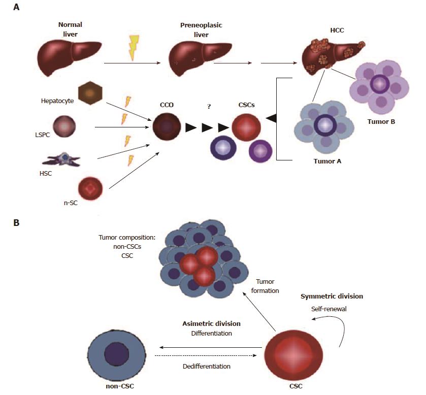
Figure 3 An integrated model of cancer cell of origin and cancer stem cell hypotheses in hepatocellular carcinoma.
A: Different cells involved in adult liver regeneration are potential targets of malignant transformation when they received hard insults (Yellow ray, i.e. carcinogenic agent, partial hepatectomy). These cells could be considered CCO (Hepatocytes, LSPC, n-SC, HSC), however many studies point to the fact that during severe or chronic damage and consistently in the beginning of HCC, hepatocytes have the main role. The relationship between CCO and CSC is not clear until now. CSCs composition is heterogeneous in HCC tumors. B: The hierarchy hypothesis shows the tumor composition: CSCs and non-CSCs. This includes the plasticity model, which indicates the interconversion by differentiation/dedifferentiation balance between non-CSCs and CSCs. LSPC: Liver stem progenitor cell; n-SC: Normal stem cell; HSC: Hepatic stellar cells.
- Citation: Flores-Téllez TN, Villa-Treviño S, Piña-Vázquez C. Road to stemness in hepatocellular carcinoma. World J Gastroenterol 2017; 23(37): 6750-6776
- URL: https://www.wjgnet.com/1007-9327/full/v23/i37/6750.htm
- DOI: https://dx.doi.org/10.3748/wjg.v23.i37.6750


