Copyright
©The Author(s) 2017.
World J Gastroenterol. Sep 28, 2017; 23(36): 6741-6746
Published online Sep 28, 2017. doi: 10.3748/wjg.v23.i36.6741
Published online Sep 28, 2017. doi: 10.3748/wjg.v23.i36.6741
Figure 1 Computed tomography scan.
A: Initial computed tomography scan of the abdomen revealing a 3 cm biloma at the gallbladder fossa with bile tracking into the sub-hepatic recess; B: Multiple rim-enhancing smaller intra-abdominal collections were also present in the upper abdomen; C: The largest intra-abdominal collection was a 9.3 cm × 8.5 cm perisplenic collection.
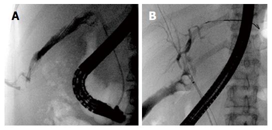
Figure 2 Endoscopic retrograde cholangiography.
A: Endoscopic retrograde cholangiography revealed a large Strasberg Type D common hepatic duct defect with contrast seen immediately in the sub-hepatic recess; B: Plastic biliary stents inserted across the biliary defect into the left and right hepatic ducts.
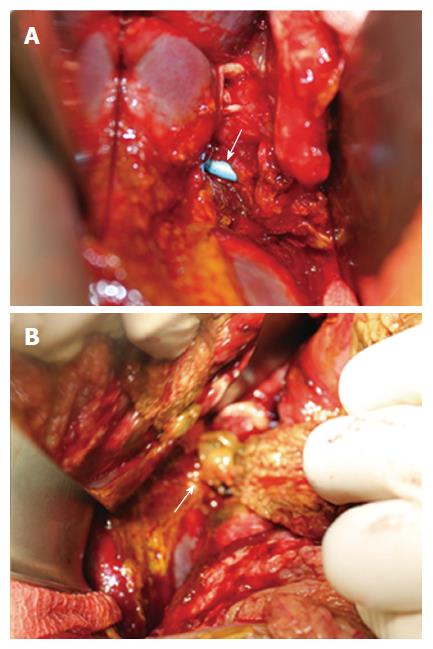
Figure 3 Damage control surgery.
A: At the time of exploratory laparotomy, endoscopically placed biliary stents were visible within a large 2 cm anterolateral common hepatic duct defect; B: A pedicled omental patch was harvested and secured to the biliary defect using absorbable sutures.
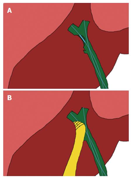
Figure 4 An illustration of the pedicled omental patch.
A: An illustration showing the location of the common hepatic duct defect and endoscopic biliary stents placed across; B: The harvested pedicled omental patch was placed over the biliary defect and secured using absorbable sutures that run through the anterior and posterior margins of the biliary defect.
- Citation: Ng JJ, Kow AWC. Pedicled omental patch as a bridging procedure for iatrogenic bile duct injury. World J Gastroenterol 2017; 23(36): 6741-6746
- URL: https://www.wjgnet.com/1007-9327/full/v23/i36/6741.htm
- DOI: https://dx.doi.org/10.3748/wjg.v23.i36.6741


