Copyright
©The Author(s) 2017.
World J Gastroenterol. Aug 7, 2017; 23(29): 5438-5450
Published online Aug 7, 2017. doi: 10.3748/wjg.v23.i29.5438
Published online Aug 7, 2017. doi: 10.3748/wjg.v23.i29.5438
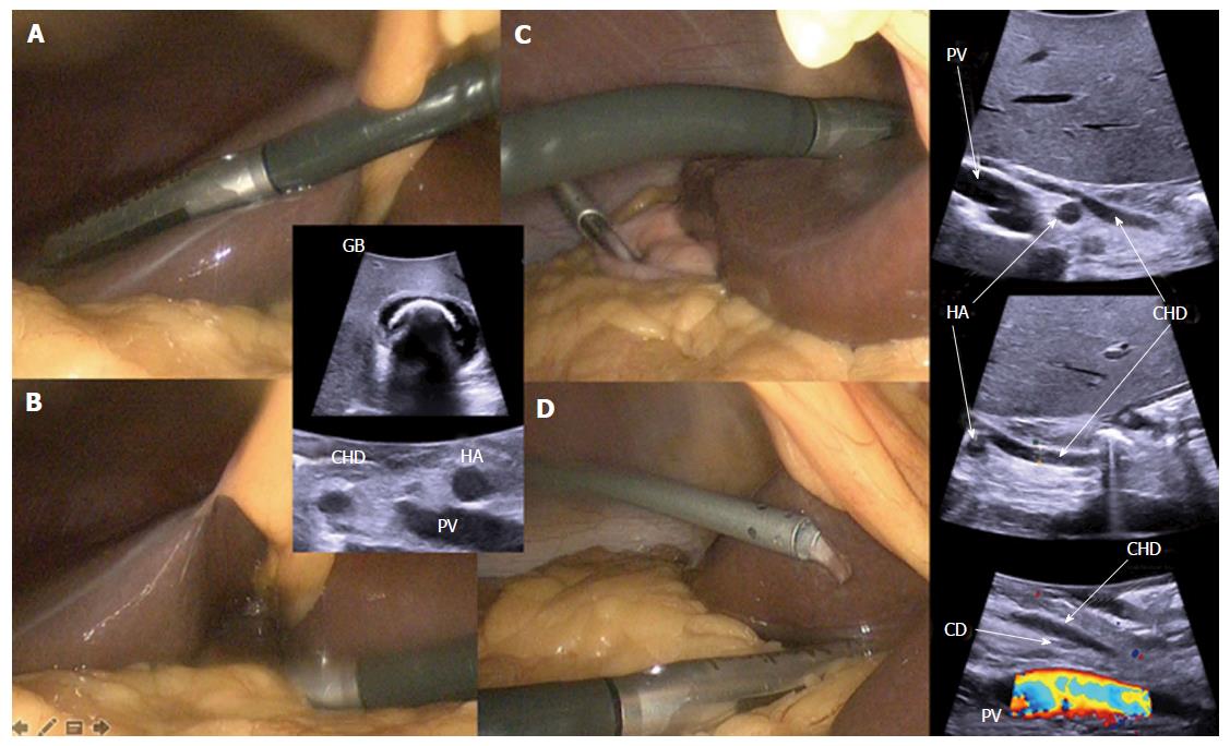
Figure 1 Laparoscopic ultrasonography: technique.
Transversal approach - A: Through the liver; B: Directly on the hepatoduodenal pedicle. Longitudinal approach - C: Through the liver; D: Directly on the hepatoduodenal pedicle (isotonic solution’s irrigation that improves acoustic coupling). CD: Cystic duct junction with the the common bile duct; CHD: Common hepatic duct; HA: Hepatic artery; PV: Portal vein; GB: Gallbladder with macrolithiasis.
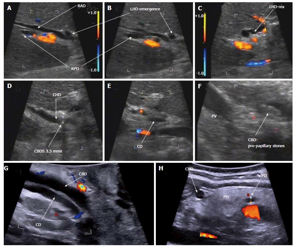
Figure 2 Laparoscopic ultrasonography: bile duct anatomy.
A-C: Biliary convergence anatomy; D-F: Classical cystic duct junction with common bile ducts stones; G and H: Intra-pancreatic bile duct. RAD: Right anterior sector duct; RPD: Right posterior sector duct; LHD: Left hepatic duct; LHD-rex: Left hepatic duct at Rex recessus; CBDS: Common bile duct stones; CD (E): Cystic duct; CHD: Common hepatic duct; CBD: Common bile duct; CD (G): Cystic duct with low implantation in the common bile duct; PD: Pancreatic duct; PH: Pancreatic head.
- Citation: Dili A, Bertrand C. Laparoscopic ultrasonography as an alternative to intraoperative cholangiography during laparoscopic cholecystectomy. World J Gastroenterol 2017; 23(29): 5438-5450
- URL: https://www.wjgnet.com/1007-9327/full/v23/i29/5438.htm
- DOI: https://dx.doi.org/10.3748/wjg.v23.i29.5438


