Copyright
©The Author(s) 2017.
World J Gastroenterol. Aug 7, 2017; 23(29): 5282-5294
Published online Aug 7, 2017. doi: 10.3748/wjg.v23.i29.5282
Published online Aug 7, 2017. doi: 10.3748/wjg.v23.i29.5282
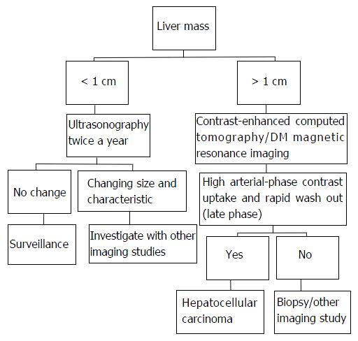
Figure 1 Imaging studies used in diagnosis, treatment planning, management and follow-up of hepatocellular carcinoma.
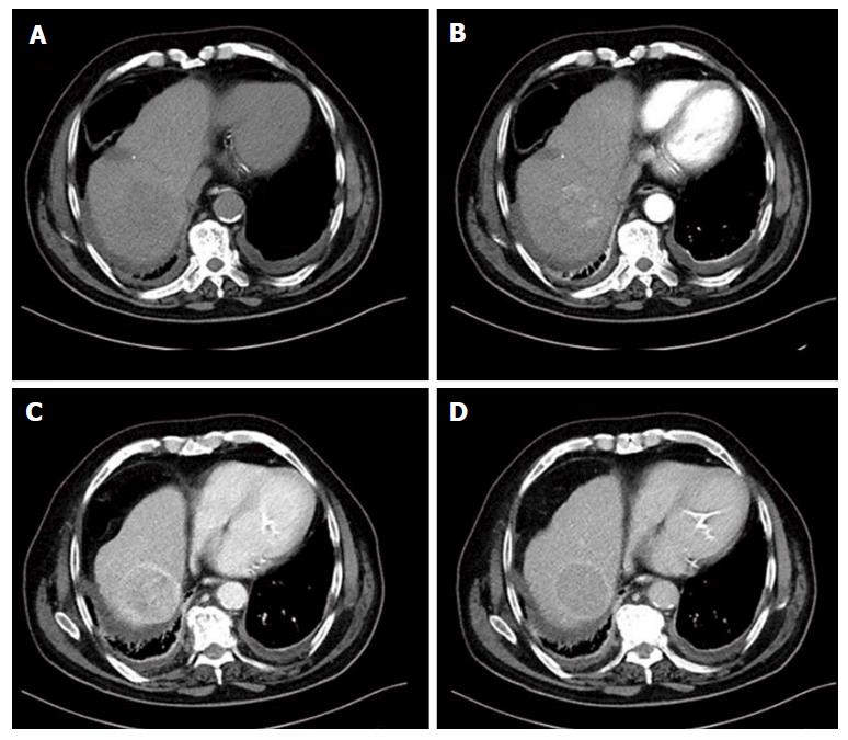
Figure 2 Multiphasic computed tomography in a large hepatocellular carcinoma located in the right liver lobe.
A: Unenhanced image; B: Lesion’s enhancement in the late hepatic arterial phase; C: Lesion’s “washout” in the portal venous phase; D: Delayed phase image. The lesion has capsule appearance most shown in the portal venous and delayed phase.
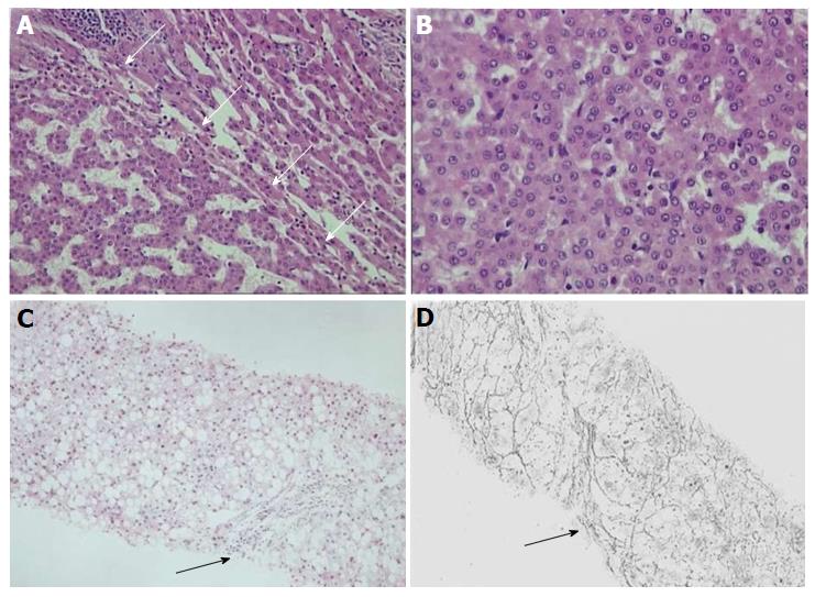
Figure 3 Well differentiated, (grade 1) hepatocellular carcinoma and early hepatocellular carcinoma with diffuse fatty change.
A: White arrows indicate the interface between HCC (left) and background liver (right); B: HCC cells show high nuclear/cytoplasmic ratio and minimal nuclear atypia. A: H/E × 100, B: H/E × 200; C and D: Early HCC with diffuse fatty change. Black arrowhead depicts a preserved portal tract. Gomori stain shows rarefaction of reticulin network. C: H/E × 100, D: Gomori stain × 100. HCC: Hepatocellular carcinoma.
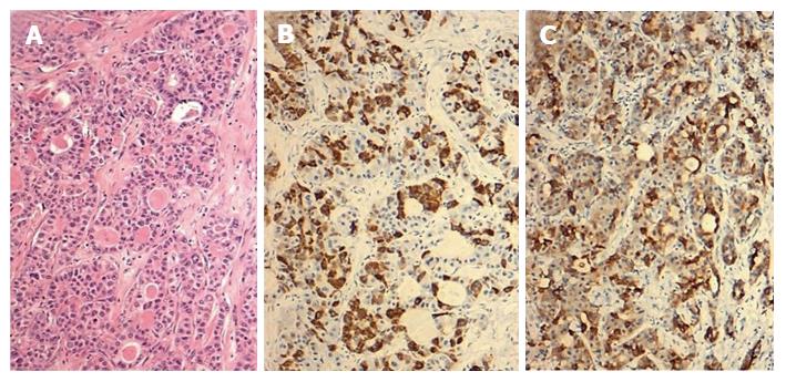
Figure 4 Combined hepatocellular-cholangiocarcinoma with stem cell features, intermediate cell subtype.
Tumor expresses both hepatocellular (HepPar1) and biliary (CK19) immunohistochemical markers. A: H/E × 100; B: HepPar1 × 100; C: CK19 × 100.
Figure 5 Intra-operative situs [prior (A) and post (B) right hepatectomy] and surgical specimen (C) of a large hepatocellular carcinoma located in the right liver lobe.
- Citation: Dimitroulis D, Damaskos C, Valsami S, Davakis S, Garmpis N, Spartalis E, Athanasiou A, Moris D, Sakellariou S, Kykalos S, Tsourouflis G, Garmpi A, Delladetsima I, Kontzoglou K, Kouraklis G. From diagnosis to treatment of hepatocellular carcinoma: An epidemic problem for both developed and developing world. World J Gastroenterol 2017; 23(29): 5282-5294
- URL: https://www.wjgnet.com/1007-9327/full/v23/i29/5282.htm
- DOI: https://dx.doi.org/10.3748/wjg.v23.i29.5282


