Copyright
©The Author(s) 2017.
World J Gastroenterol. Mar 28, 2017; 23(12): 2194-2200
Published online Mar 28, 2017. doi: 10.3748/wjg.v23.i12.2194
Published online Mar 28, 2017. doi: 10.3748/wjg.v23.i12.2194
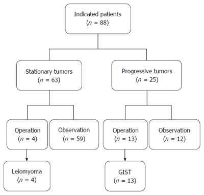
Figure 1 Flow chart of management of 88 indicated patients with submucosal tumors originating from the muscularis propria in the stomach.
EUS: Endoscopic ultrasound; GIST: Gastrointestinal stromal tumor.
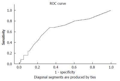
Figure 2 Receiver operating characteristic curve analysis of tumor size for predicting potential tumor progression.
Initial tumor size of 1.4 cm was determined as the optimal cut-off size, with a sensitivity of 68.0%, a specificity of 66.7%, and an accuracy of 67.0%.
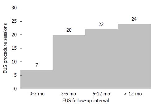
Figure 3 Intervals of endoscopic ultrasound follow-up in 25 patients with 1-3 cm gastric submucosal tumors originating from the muscularis propria in tumor progression.
EUS: Endoscopic ultrasound.
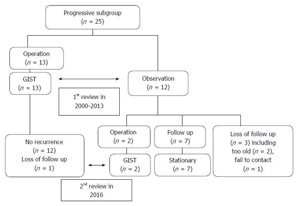
Figure 4 Flow chart of patients in the progressive subgroup.
These patients were reviewed twice; the first was based on medical records in 2013 and the second was performed by phone calls as well as based on medical records in 2016.
- Citation: Hu ML, Wu KL, Changchien CS, Chuah SK, Chiu YC. Endosonographic surveillance of 1-3 cm gastric submucosal tumors originating from muscularis propria. World J Gastroenterol 2017; 23(12): 2194-2200
- URL: https://www.wjgnet.com/1007-9327/full/v23/i12/2194.htm
- DOI: https://dx.doi.org/10.3748/wjg.v23.i12.2194


