Copyright
©The Author(s) 2017.
World J Gastroenterol. Jan 7, 2017; 23(1): 25-41
Published online Jan 7, 2017. doi: 10.3748/wjg.v23.i1.25
Published online Jan 7, 2017. doi: 10.3748/wjg.v23.i1.25
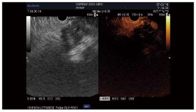
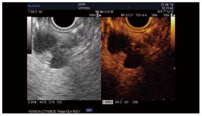
Figure 1 Contrast-enhanced harmonics-endoscopic ultrasound of pancreatic adenocarcinoma.
Standard endoscopic ultrasound image (left) of a hypoenhanced lesion of the pancreatic body. The contrast-enhanced image (right) shows a hypoenhanced adenocarcinoma. The dilated pancreatic duct is seen upstream of the lesion.
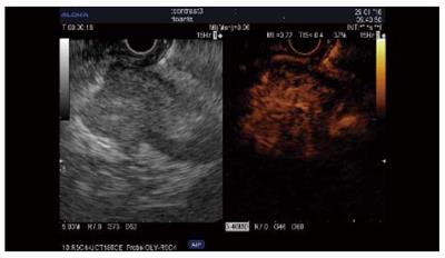
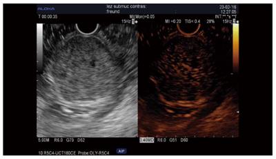
Figure 2 Contrast-enhanced harmonics-endoscopic ultrasound of a neuroendocrine pancreatic tumor.
Standard endoscopic ultrasound image (left) of a hypoenhanced, well-delineated lesion of the head of the pancreas. The contrast image (right) shows a hyperenhanced lesion that is suggestive of a neuroendocrine tumor, as later proved by fine needle aspiration.
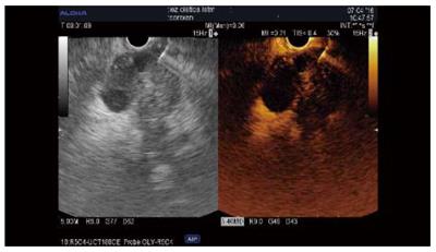
Figure 3 Contrast-enhanced harmonics-endoscopic ultrasound-fine needle aspiration of solid pancreatic adenocarcinoma.
Standard endoscopic ultrasound view of a hypoenhanced, inhomogenous adenocarcinoma of the head of the pancreas, showing anechoic parts suggestive of necrosis. The contrast image (right) highlights these avascular parts of the lesion, and the needle inside is clearly seen as avoiding them.
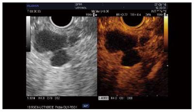
Figure 4 Contrast-enhanced harmonics-endoscopic ultrasound in mucinous cystadenomashowing features suggestive of malignancy.
Standard endoscopic ultrasound image (left) of a macrocystic lesion of the pancreas with mural nodules and thin septae inside. The contrast image (right) shows vascularized mural nodules and no contrast uptake in some of the septae.
Figure 5 CH-endoscopic ultrasound-fine needle aspiration of a vascularized mural nodules within mucinous cystadenoma.
An endoscopic ultrasound-fine needle aspiration needle during the puncture of one of the mural nodules is better seen on the contrast image (right).
Figure 6 Contrast-enhanced harmonics endoscopic ultrasound of gastric gastrointestinal stromal tumor.
Endoscopic ultrasound standard image (right) of a gastric gastrointestinal stromal tumor of the muscularis propria. The contrast image (left) shows hyperenhancement of the lesion.
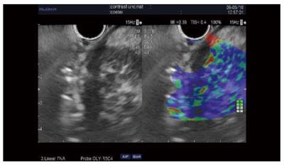
Figure 7 Elastography of pancreatic neuroendocrine tumor - qualitative assessment.
Endoscopic ultrasound standard image (right) showing a well-delineated hypoenhanced lesion of the head of the pancreas. The elastography image (left) exhibits a blue pattern in this neuroendocrine tumor.
- Citation: Seicean A, Mosteanu O, Seicean R. Maximizing the endosonography: The role of contrast harmonics, elastography and confocal endomicroscopy. World J Gastroenterol 2017; 23(1): 25-41
- URL: https://www.wjgnet.com/1007-9327/full/v23/i1/25.htm
- DOI: https://dx.doi.org/10.3748/wjg.v23.i1.25


