Copyright
©The Author(s) 2016.
World J Gastroenterol. Feb 14, 2016; 22(6): 2081-2091
Published online Feb 14, 2016. doi: 10.3748/wjg.v22.i6.2081
Published online Feb 14, 2016. doi: 10.3748/wjg.v22.i6.2081
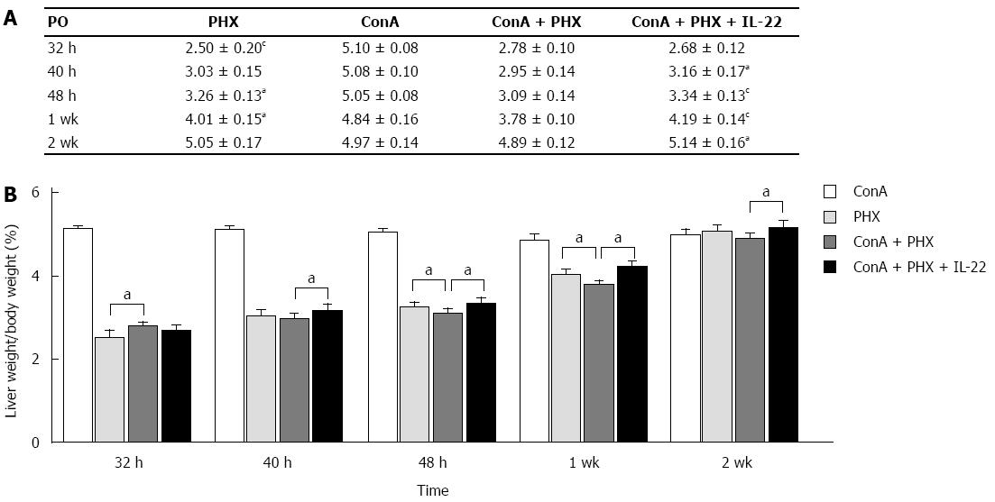
Figure 1 Liver weight/body weight ratios following partial hepatectomy.
A and B: Increases in the liver weight/body weight ratio were observed in the PHX, concanavalin A (ConA) + PHX and ConA + PHX + interleukin (IL)-22 groups, and all groups returned to normal liver weights by 2 wk. Compared with the ConA + PHX group, the ConA + PHX + IL-22 group exhibited greater increases that reached significance at 40 h, 48 h, 1 wk and 2 wk (aP < 0.05); these differences were particularly notable at 48 h and 1 wk (cP < 0.01). The increases in the PHX group were significantly different from those in the ConA + PHX group at 48 h and 1 wk (aP < 0.05); however, the increase in the PHX group at 32 h was less than that in the ConA + PHX group (cP < 0.01). These data were correlated with the cellular swelling in the liver at 32 h in the ConA + PHX group.
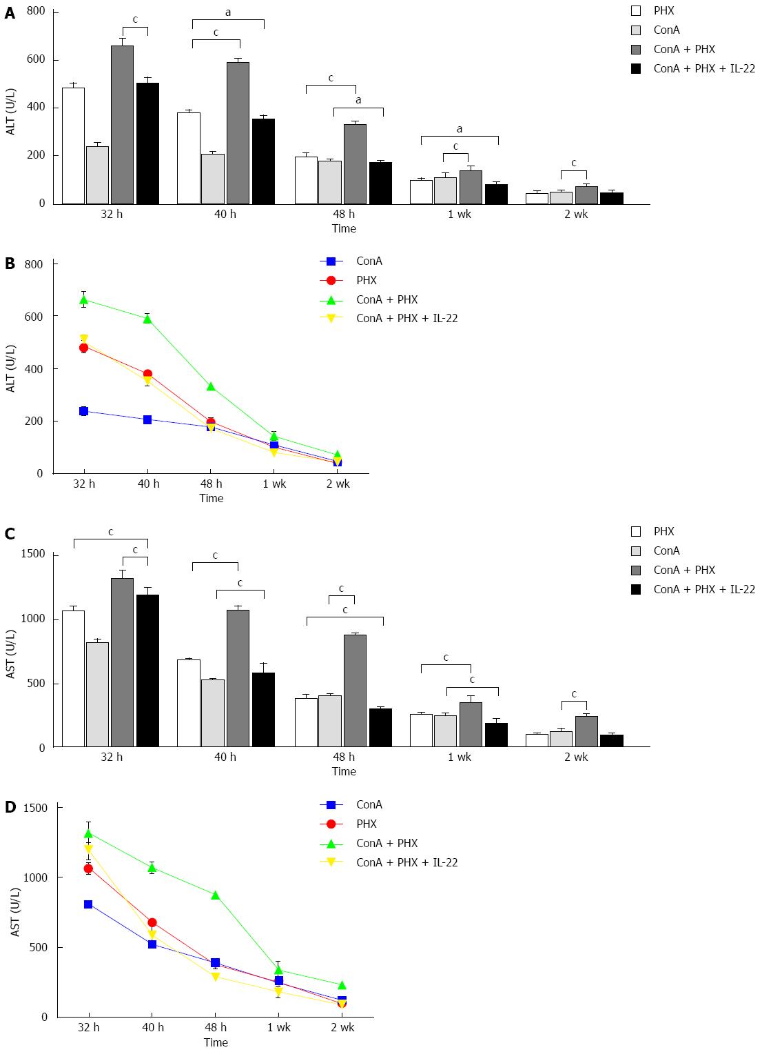
Figure 2 Serum alanine aminotransferase and aspartate aminotransferase levels after partial hepatectomy.
A-D: Decreases in serum alanine aminotransferase (ALT) and aspartate aminotransferase (AST) were observed after 32 h, particularly from 40 h to 1 wk. Compared to the concanavalin A (ConA) + PHX group, the serum ALT and AST levels of the ConA + PHX + interleukin (IL)-22 group decreased, and the difference were significant at all time points (cP < 0.01). Furthermore, with the recombinant IL-22 pretreatment, the ALT and AST levels in the ConA + PHX + IL-22 group decreased to significantly greater extents than the levels in the ConA and PHX groups at 40 h, 48 h and 1 wk (aP < 0.05).
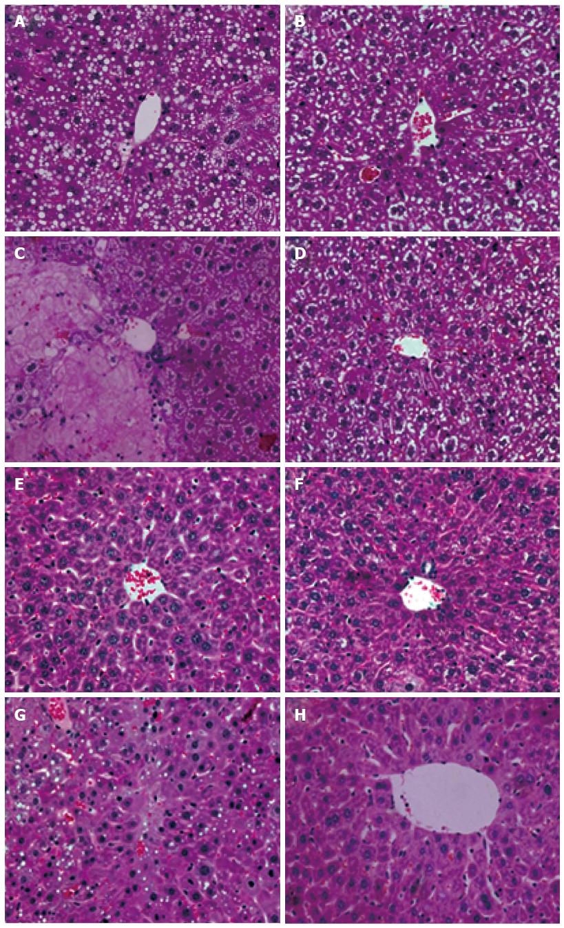
Figure 3 Representative hematoxylin and eosin staining of the remnant livers after partial hepatectomy.
A-D: The PHX, concanavalin A (ConA), ConA + PHX and ConA + PHX + interleukin (IL)-22 groups, respectively, at 48 h after partial hepatectomy; E-H: The PHX, ConA, ConA + PHX and ConA + PHX + IL-22 groups, respectively, at 2 wk after partial hepatectomy.
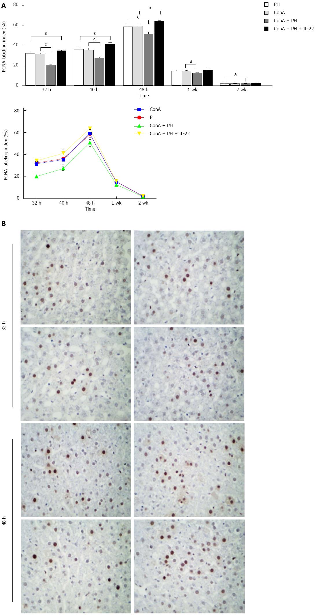
Figure 4 Proliferating cell nuclear antigen labeling indices in the remnant livers after partial hepatectomy.
A: Compared with the concanavalin A (ConA) + PHX group, the proliferating cell nuclear antigen (PCNA) labeling indices were significantly increased in the ConA, PHX and ConA + PHX + interleukin (IL)-22 groups at all of the time points (aP < 0.05), particularly at 32, 40, and 48 h (cP < 0.01). Furthermore, the increases in the PCNA levels in the ConA + PHX + IL-22 group were significantly greater than those in the ConA and PHX groups at 32 h, 40 h and 48 h (aP < 0.05); B: PCNA labeling of the cells at 32 h and 48 h after hepatectomy in the PHX, ConA, ConA + PHX and ConA + PHX + IL-22 groups, respectively.
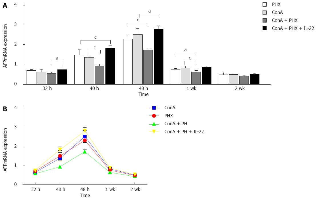
Figure 5 Hepatic alpha fetal protein mRNA expression in the remnant liver after partial hepatectomy.
A, B: Alpha fetal protein (AFP) mRNA expression began to increase at 32 h and significantly increased to peak at 48 h after hepatectomy. With the interleukin (IL)-22 pretreatment, the AFP mRNA levels significantly increased at 32 h after partial hepatectomy compared with the concanavalin A (ConA) + PHX group (aP < 0.05), and marked increases were observed at 40 h, 48 h and 1 wk (cP < 0.01). However, the difference was not significant at 2 wk (P > 0.05).
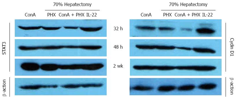
Figure 6 Western blot detection of the activation of STAT3 and Cyclin D1 in the remnant livers after partial hepatectomy.
With the administration of interleukin (IL)-22, STAT3 and Cyclin D1 were activated much earlier, and the most significant increases in the four groups were observed at 32 h, 48 h, and 2 wk; whereas the least significant increases were observed in the concanavalin A (ConA) + PHX group at all of the time points.
- Citation: Zhang YM, Liu ZR, Cui ZL, Yang C, Yang L, Li Y, Shen ZY. Interleukin-22 contributes to liver regeneration in mice with concanavalin A-induced hepatitis after hepatectomy. World J Gastroenterol 2016; 22(6): 2081-2091
- URL: https://www.wjgnet.com/1007-9327/full/v22/i6/2081.htm
- DOI: https://dx.doi.org/10.3748/wjg.v22.i6.2081


