Copyright
©The Author(s) 2016.
World J Gastroenterol. Feb 7, 2016; 22(5): 1767-1778
Published online Feb 7, 2016. doi: 10.3748/wjg.v22.i5.1767
Published online Feb 7, 2016. doi: 10.3748/wjg.v22.i5.1767
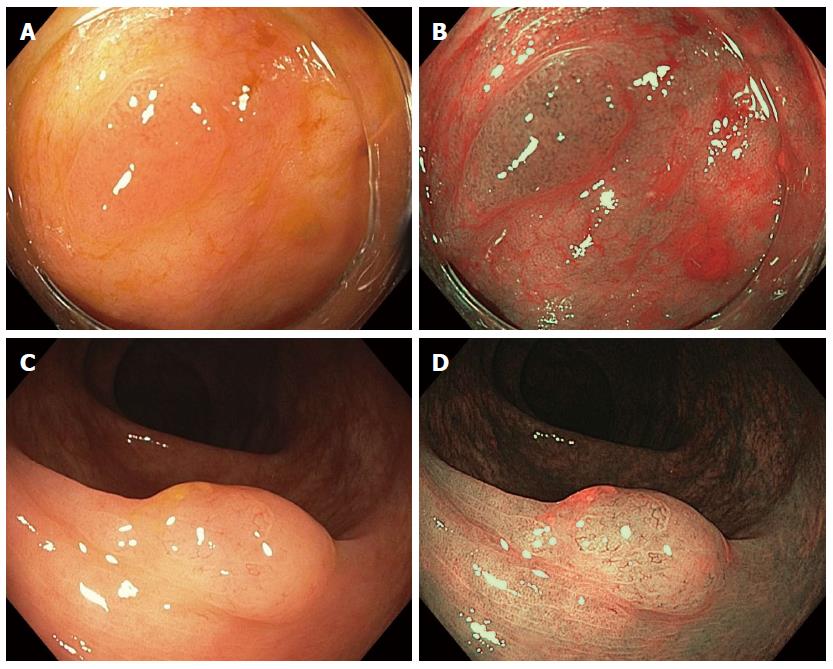
Figure 1 Tubular adenoma and small sessile serrated adenoma under white light (A, C) and narrow-band imaging (B, D), respectively.
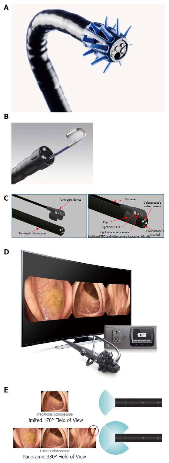
Figure 2 Endoscuff (A), Third-Eye retroscope (B), Third-Eye panoramic (C), full spectrum endoscopy (D: Endoscope, processor and image; E: Endoscopic view).
- Citation: Aranda-Hernández J, Hwang J, Kandel G. Seeing better - Evidence based recommendations on optimizing colonoscopy adenoma detection rate. World J Gastroenterol 2016; 22(5): 1767-1778
- URL: https://www.wjgnet.com/1007-9327/full/v22/i5/1767.htm
- DOI: https://dx.doi.org/10.3748/wjg.v22.i5.1767


