Copyright
©The Author(s) 2016.
World J Gastroenterol. Dec 7, 2016; 22(45): 10038-10044
Published online Dec 7, 2016. doi: 10.3748/wjg.v22.i45.10038
Published online Dec 7, 2016. doi: 10.3748/wjg.v22.i45.10038
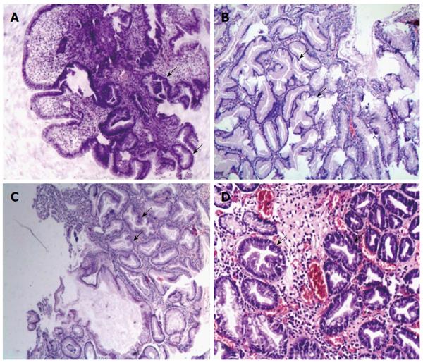
Figure 1 Typical pathological images of serrated lesions in upper gastrointestinal tract.
Serrated lesions characterized by epithelial cells with luminal infolding and a serrated growth pattern were shown. A: Serrated hyperplasia in esophagitis (× 40); B: Serrated hyperplasia in the Ménétrier gastropathy: marked foveolar hyperplasia and glandular cysts with serrated lesions in the stomach (× 100); C: Hyperplastic polyp in the stomach: a serrated polyp without overt cytological atypia showed narrowed crypt bases that were predominantly lined with immature cells (× 100); D: Serrated adenoma with low grade dysplasia in the duodenum: a serrated polyp with enlarged nuclei, a pencil-shaped, hyperchromaticity and nuclear stratification (× 100).
- Citation: Cao HL, Dong WX, Xu MQ, Zhang YJ, Wang SN, Piao MY, Cao XC, Wang BM. Clinical features of upper gastrointestinal serrated lesions: An endoscopy database analysis of 98746 patients. World J Gastroenterol 2016; 22(45): 10038-10044
- URL: https://www.wjgnet.com/1007-9327/full/v22/i45/10038.htm
- DOI: https://dx.doi.org/10.3748/wjg.v22.i45.10038


