Copyright
©The Author(s) 2016.
World J Gastroenterol. Jan 28, 2016; 22(4): 1357-1366
Published online Jan 28, 2016. doi: 10.3748/wjg.v22.i4.1357
Published online Jan 28, 2016. doi: 10.3748/wjg.v22.i4.1357
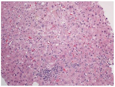
Figure 1 Liver biopsy with cholestasis, mild portal and lobular inflammation, and apoptotic bodies.
Hematoxylin and eosin stain, magnification × 100.
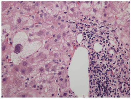
Figure 2 Lymphocyte predominant portal inflammation and a damaged bile duct (Poulsen Christofferson lesion).
Periportal hepatocytes show ballooning/feathery degeneration. Hematoxylin and eosin stain, magnification × 200.
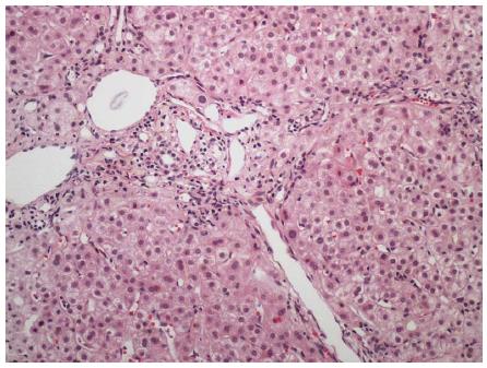
Figure 3 Portal and periportal fibrosis in a patient with concurrent human immunodeficiency virus and acute hepatitis C infection.
Hematoxylin and eosin stain, magnification × 100.
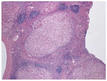
Figure 4 Fibrous septa with lymphocyte predominant inflammatory infiltrates and multiple lymphoid aggregates, in a patient with cirrhosis secondary to hepatitis C.
Hematoxylin and eosin stain, magnification × 40.
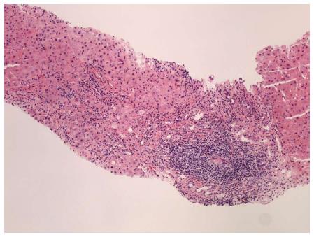
Figure 5 Portal lymphoid aggregate and lymphoplasmacytic inflammation with severe interface hepatitis, indicative of an overlap syndrome of autoimmune hepatitis and hepatitis C.
Hematoxylin and eosin stain, magnification × 100.
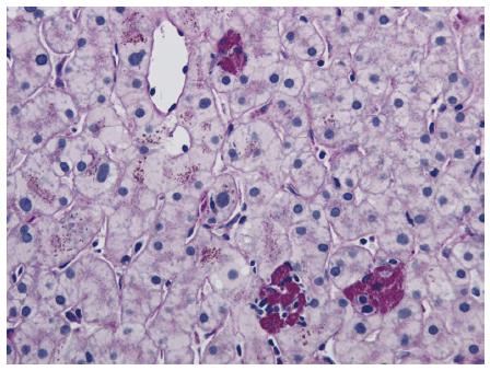
Figure 6 Small clusters of lobular macrophages containing periodic acid Schiff positive-diastase resistant cytoplasmic debris.
Periodic acid Schiff with diastase stain, magnification × 200.
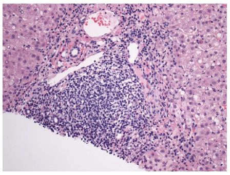
Figure 7 Portal lymphoid aggregate and lymphoplasmacytic inflammation with interface hepatitis and occasional apoptotic hepatocyte at the portal-periportal interface.
Hematoxylin and eosin stain, magnification × 200.
Figure 8 Portal tract with lymphoplasmacytic inflammation, and enclosing small clusters of hepatocytes indicative of recent episode of active hepatitis.
Hematoxylin and eosin stain, magnification × 200.
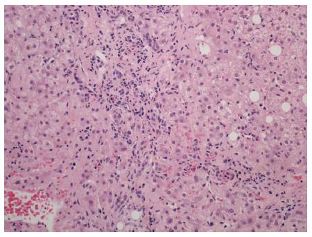
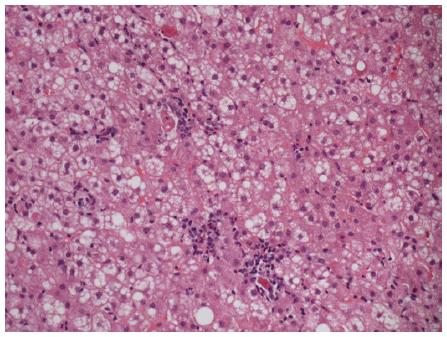
Figure 9 Spotty necrosis characterized by small foci of lobular necroinflammation and associated apoptotic hepatocytes.
Hematoxylin and eosin stain, magnification × 200.
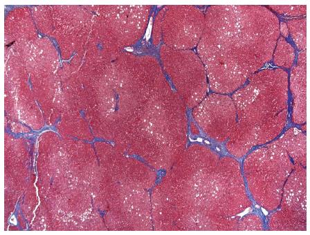
Figure 10 Cirrhosis characterized by bridging fibrosis with nodule formation.
Masson’s trichrome stain, magnification × 40.
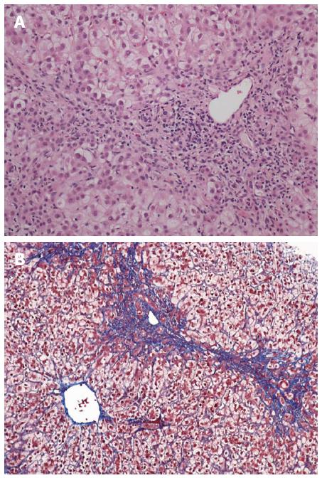
Figure 11 Fibrosing cholestatic hepatitis with mixed portal inflammation, bile duct damage, interface hepatitis, ductular reaction and fibrosis.
Hematoxylin and eosin stain, magnification × 100 (A); and extensive pericellular and perisinusoidal fibrosis (B; Trichrome stain, magnification × 100).
- Citation: Dhingra S, Ward SC, Thung SN. Liver pathology of hepatitis C, beyond grading and staging of the disease. World J Gastroenterol 2016; 22(4): 1357-1366
- URL: https://www.wjgnet.com/1007-9327/full/v22/i4/1357.htm
- DOI: https://dx.doi.org/10.3748/wjg.v22.i4.1357


