Copyright
©The Author(s) 2016.
World J Gastroenterol. Oct 7, 2016; 22(37): 8257-8270
Published online Oct 7, 2016. doi: 10.3748/wjg.v22.i37.8257
Published online Oct 7, 2016. doi: 10.3748/wjg.v22.i37.8257
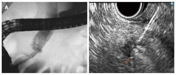
Figure 1 Endobiliary and pancreatic radiofrequency ablation.
A: Fluoroscopy: The Habib TM EndoHBP inside the common bile duct; B: EUS: The EUSRA RF electrode (STARmed) inserted in a PNET. Tip of the probe (orange arrow). EUS: Endoscopic ultrasonography; RF: Radiofrequency.
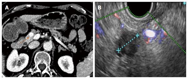
Figure 2 Ten millimeter pancreatic neuroendocrine tumour before radiofrequency ablation.
Chromogranin at diagnosis: 239 ng/mL. A: The corresponding CT image (orange arrow); B: The corresponding Doppler endoscopic ultrasonography image.
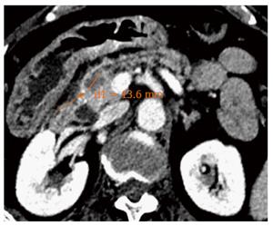
Figure 3 Same pancreatic neuroendocrine tumour showing necrosis (orange arrow) two days after Radiofrequency ablation.
Chromogranin level decreased to 36 ng/mL.
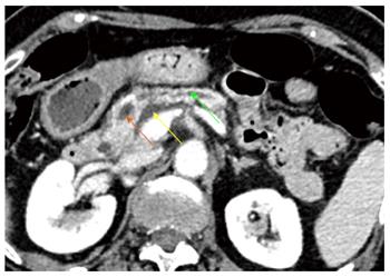
Figure 4 Patient suffered a mild acute pancreatitis 3 wk after radiofrequency ablation.
CT revealed a peripancreatic fluid collection (yellow arrow) and tumour necrosis (orange arrow) with slight dilation of the upstream pancreatic duct (green arrow).
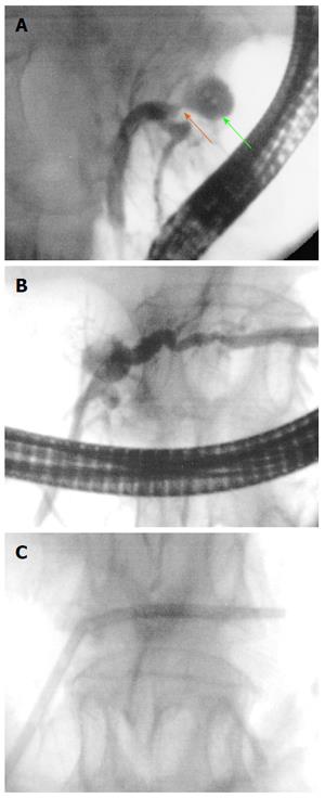
Figure 5 Pancreatic duct stenosis after pancreatic radiofrequency ablation.
A and B: ERCP revealed a necrotic cavity (green arrow) and a pancreatic duct stenosis (orange arrow); C: A plastic stent was inserted.
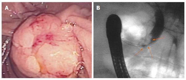
Figure 6 Ampullary adenoma with bile duct extension.
A: Endoscopic image of a large lesion involving the ampulla and the adjacent duodenum; B: Bile duct extension at endoscopic retrograde cholangiopancreatography (orange arrows).
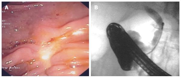
Figure 7 Results after endoscopic papillectomy and biliary radiofrequency ablation at two-year follow-up.
A: Endoscopy showed complete resection without tumour recurrence; B: No longer bile duct ingrowth at endoscopic retrograde cholangiopancreatography. Biopsies were negative repeatedly.
- Citation: Alvarez-Sánchez MV, Napoléon B. Review of endoscopic radiofrequency in biliopancreatic tumours with emphasis on clinical benefits, controversies and safety. World J Gastroenterol 2016; 22(37): 8257-8270
- URL: https://www.wjgnet.com/1007-9327/full/v22/i37/8257.htm
- DOI: https://dx.doi.org/10.3748/wjg.v22.i37.8257


