Copyright
©The Author(s) 2016.
World J Gastroenterol. Jul 14, 2016; 22(26): 6089-6094
Published online Jul 14, 2016. doi: 10.3748/wjg.v22.i26.6089
Published online Jul 14, 2016. doi: 10.3748/wjg.v22.i26.6089
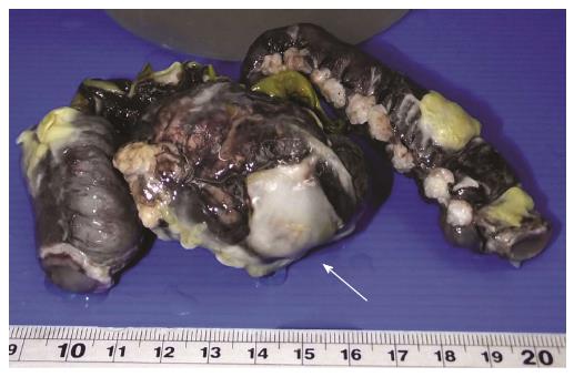
Figure 1 Specimen after fixation revealed a perforated ulcer with fibrofibrinous exudate (arrow).
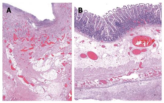
Figure 2 Histopathologic findings (hematoxylin-eosin staining, magnification × 40).
A: Base of ulcer revealed inflammation, granulation tissue and fibrosis; B: Remaining ileum showed lymphoid follicle depletion, submucosal and serosal congestion with diffuse peritonitis.
- Citation: Lerkvaleekul B, Treepongkaruna S, Saisawat P, Thanachatchairattana P, Angkathunyakul N, Ruangwattanapaisarn N, Vilaiyuk S. Henoch-Schönlein purpura from vasculitis to intestinal perforation: A case report and literature review. World J Gastroenterol 2016; 22(26): 6089-6094
- URL: https://www.wjgnet.com/1007-9327/full/v22/i26/6089.htm
- DOI: https://dx.doi.org/10.3748/wjg.v22.i26.6089


