Copyright
©The Author(s) 2016.
World J Gastroenterol. Jul 14, 2016; 22(26): 5917-5926
Published online Jul 14, 2016. doi: 10.3748/wjg.v22.i26.5917
Published online Jul 14, 2016. doi: 10.3748/wjg.v22.i26.5917
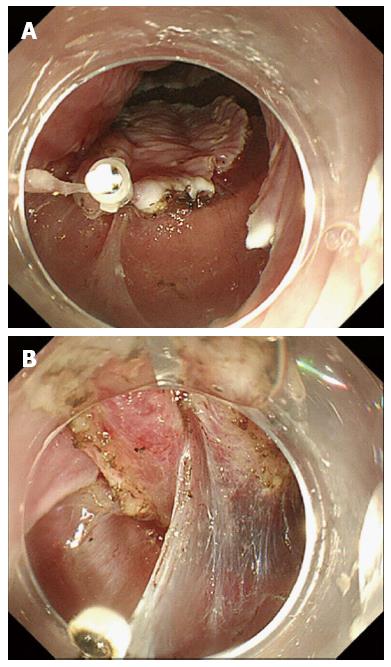
Figure 1 Clip with the line was placed on the lesion (A), and the clip and line method facilitated adequate traction (B).
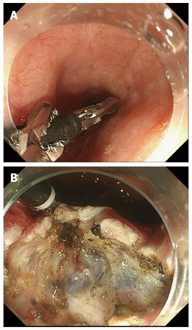
Figure 2 External grasping forceps was carefully delivered with the help of the another grasping forceps (A) and the lesion was grasped by the external grasping forceps (B).
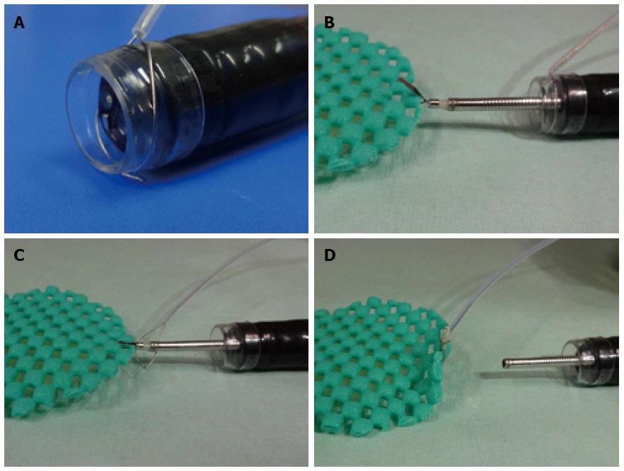
Figure 3 Schematic of our clip and snare method using a pre-looping technique.
A: Endoscope with the prelooped snare; B: The clip was inserted through the working channel of the endoscope and used to grasp the mucosal flap on one side of the lesion; C: The snare, which had been pre-looped over the scope, was loosened and moved along the forceps up to the clip. We tightened the snare to grasp the clip; D: We were then able to release the clip from the forceps (from Ota et al[19]).
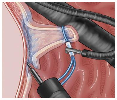
Figure 4 Schematic view of internal traction method (from Chen et al[22]).
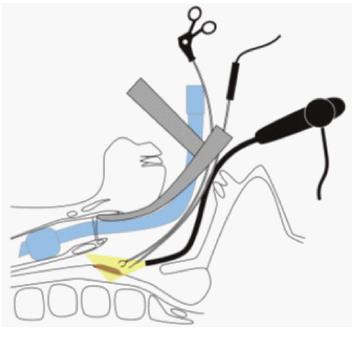
Figure 5 Schematic view of double-scope method (from Fujii et al[25]).
Figure 6 Fraenkel laryngeal forceps (A) and the lesion was grasped by the Fraenkel laryngeal forceps (B).
Figure 7 Schematic view and surgical setup of endoscopic laryngopharyngeal surgery (from Tateya et al[35]).
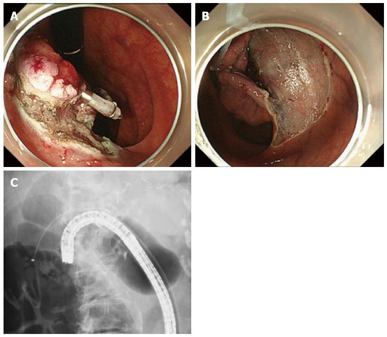
Figure 8 Clip-and-snare method enabled pushing and pulling movements for traction.
A: The lesion was pulled anally in retroflexion view using a balloon overtube in the deep colon; B: Pushing the snare to facilitate visualization of submucosal layer; C: Traction is maintained by the snare and the clip independent of the scope.
- Citation: Tsuji K, Yoshida N, Nakanishi H, Takemura K, Yamada S, Doyama H. Recent traction methods for endoscopic submucosal dissection. World J Gastroenterol 2016; 22(26): 5917-5926
- URL: https://www.wjgnet.com/1007-9327/full/v22/i26/5917.htm
- DOI: https://dx.doi.org/10.3748/wjg.v22.i26.5917


