Copyright
©The Author(s) 2016.
World J Gastroenterol. Jul 7, 2016; 22(25): 5694-5717
Published online Jul 7, 2016. doi: 10.3748/wjg.v22.i25.5694
Published online Jul 7, 2016. doi: 10.3748/wjg.v22.i25.5694
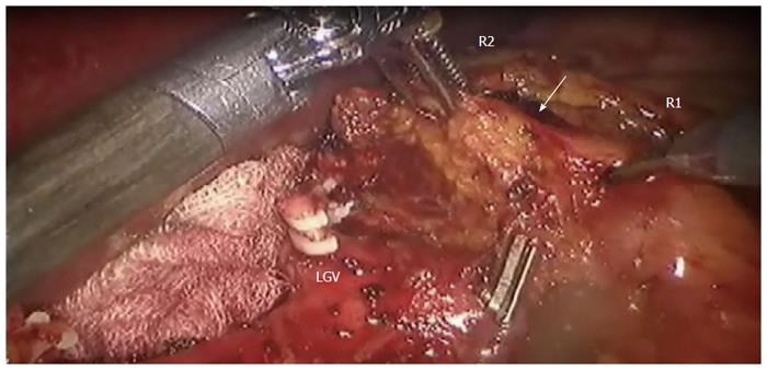
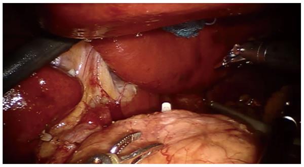
Figure 1 Adipose tissue including the station No.
8a lymph nodes (white arrow) is pulled up by the 2nd robotic (R2) arm dissected by the 1st robotic (R1) arm. Clipped on Hem-o-lock is Left gastric vein (LGV). Provided by Roviello F, University of Siena.
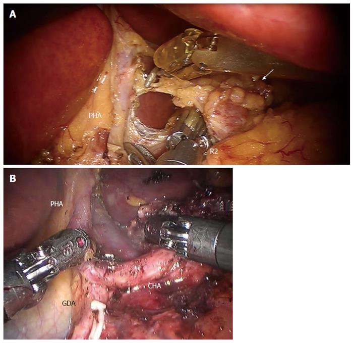
Figure 2 Dissection of No.
8 lymph nodes (white arrow) continues medially, through the traction of the 2nd robotic arm (R2), exposing (A) the proper hepatic artery and (B) the common hepatic artery. Provided by A: Coratti A, University of Florence; B: Patriti A. USL2 Spoleto. PHA: Proper hepatic artery; CHA: Common hepatic artery; GDA: Gastroduodenal artery.
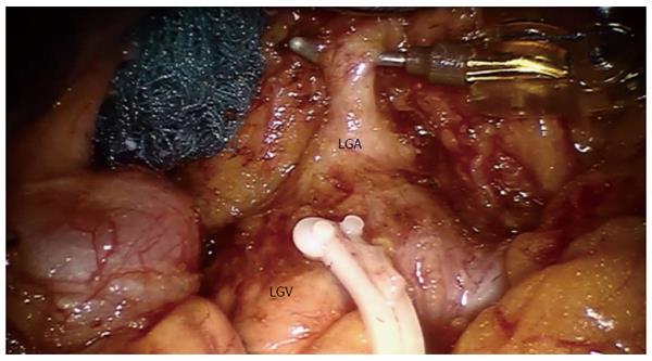
Figure 3 Exposition of left gastric artery after No.
7 lymph nodes dissection. Clipped on Hem-o-lock is the left gastric vein (LGV). Provided by Coratti A, University of Florence. LGA: Left gastric artery.
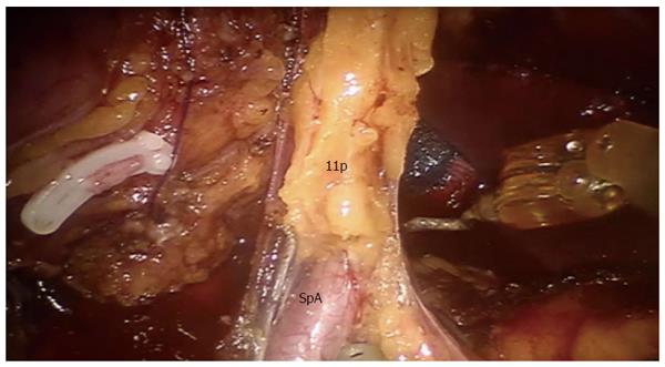
Figure 4 Dissection of 11p lymph nodes.
Provided by Coratti A, University of Florence. SpA: Splenic artery.
Figure 5 Exposure of the supra pancreatic area after supra pancreatic lymph nodes dissection.
Provided by Coratti A, University of Florence.
- Citation: Caruso S, Patriti A, Roviello F, De Franco L, Franceschini F, Coratti A, Ceccarelli G. Laparoscopic and robot-assisted gastrectomy for gastric cancer: Current considerations. World J Gastroenterol 2016; 22(25): 5694-5717
- URL: https://www.wjgnet.com/1007-9327/full/v22/i25/5694.htm
- DOI: https://dx.doi.org/10.3748/wjg.v22.i25.5694


