Copyright
©The Author(s) 2016.
World J Gastroenterol. May 21, 2016; 22(19): 4781-4785
Published online May 21, 2016. doi: 10.3748/wjg.v22.i19.4781
Published online May 21, 2016. doi: 10.3748/wjg.v22.i19.4781
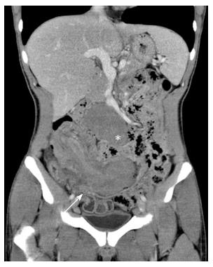
Figure 1 Contrast-enhanced computed tomography showed a well-defined homogeneous mass (asterisk) in the mesenteric root region, together with a long segmental wall thickening in the ileum with ileocolic-type intussusception (arrow).
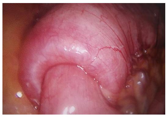
Figure 2 Invagination of the terminal ileum into the proximal colon.
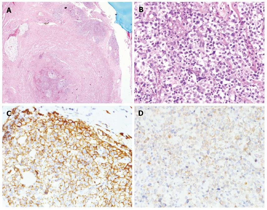
Figure 3 Histological and immunohistological examination of the specimens showing diffuse large B-cell non-Hodgkin's lymphoma.
A: Magnification × 20; HE staining. Tumor cell infiltrates can be observed in the serosal layer of the appendix; B: Magnification × 400, HE staining, polymorphic large B cells; C: Magnification × 400, CD20(+); D: Magnification × 400, Bcl-2(+). Bcl-2: B-cell lymphoma 2; HE: Hematoxylin and eosin.
- Citation: Yang TW, Lin YY, Tsuei YW, Chen YL, Huang CY, Hsu SD. Successful management of adult lymphoma-associated intussusception by laparoscopic reduction and appendectomy. World J Gastroenterol 2016; 22(19): 4781-4785
- URL: https://www.wjgnet.com/1007-9327/full/v22/i19/4781.htm
- DOI: https://dx.doi.org/10.3748/wjg.v22.i19.4781


